

Abdominal imaging addresses the health of all organs in your abdomen and pelvis, including the liver, gall bladder, bile ducts, spleen, pancreas, adrenal glands, kidneys, stomach, small and large intestine, aorta, inferior vena cava, male/female pelvic organs, bones and more.
Some Abdominal Imaging Procedures Include:
This is an accurate and noninvasive imaging procedure used to assess and evaluate certain gastrointestinal problems, such as inflammatory bowel disease (including Crohn’s Disease), infectious enteritis, lymphoma or tuberculosis. It also can be used in patients with gastrointestinal bleeding to determine if a small bowel polyp is causing the bleeding. Enterography may be performed using MRI or CT.
Ultrasound imaging of the pelvis uses sound waves to produce pictures of the structures and organs in the lower abdomen and pelvis. There are various types of pelvic ultrasound including abdominal, vaginal and rectal. These exams are frequently used to evaluate the reproductive and urinary systems. Ultrasound is safe, noninvasive and does not use ionizing radiation.
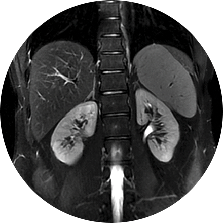
A renal ultrasound is a safe and painless test that uses sound waves to produce images of the kidneys and bladder. The kidneys are a pair of bean-shaped organs located toward the back of the abdominal cavity, just above the waist. They remove waste products from the blood and produce urine.