

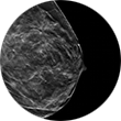


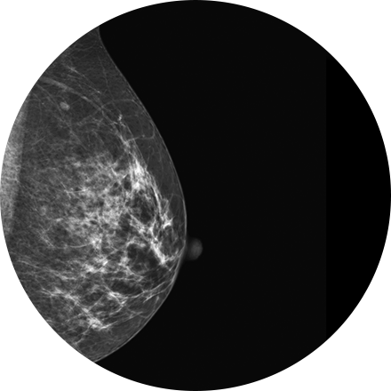
3D mammography (tomosynthesis) is an FDA-approved technology that complements conventional 2D mammography. While traditional mammography generates 2D images, 3D mammography creates multiple thin 3D image slices that allow each section of the breast tissue to be seen more clearly.
A 3D mammogram looks and feels like a regular mammogram. An x-ray arm scans over your breast taking multiple 3D images at various angles. Each scan takes about 4 seconds and the entire 3D mammogram lasts about 15-20 minutes.
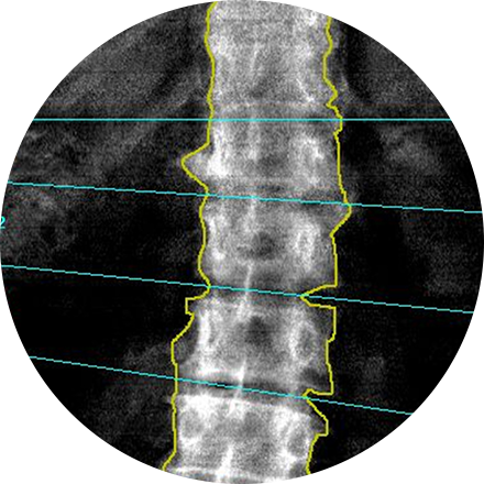
A dual-energy X-ray absorptiometry (DEXA) scan measures the density and mineral content in bone, most often in the hip or lower spine. It is the most accurate method of determining bone density and potential problems related to bone loss. This test is a valuable tool for diagnosing osteoporosis, which often has no symptoms until you suffer a fracture. A bone density scan can diagnose the disease at its earliest stages, which means you can begin receiving treatment to protect your bones sooner.
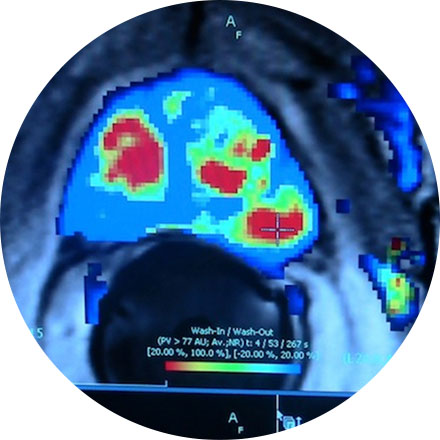
Some women — because of their family history, a genetic tendency or certain other factors — should be screened with MRI in addition to mammograms. The number of women who fall into this category is small: less than 2 percent of all the women in the United States. Talk with your doctor about your history and whether you should have additional tests.
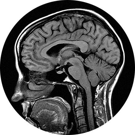
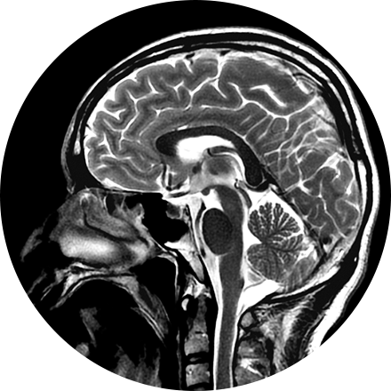
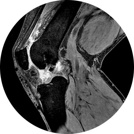
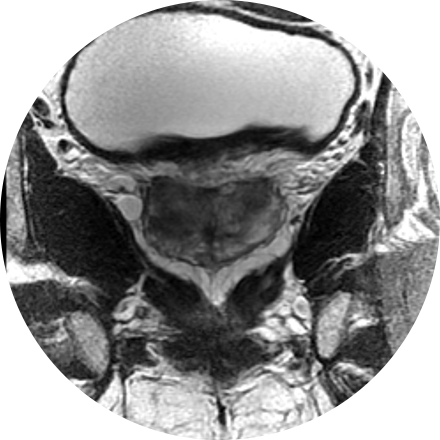
Breast MRI helps to detect small lesions sometimes missed by mammography — without using radiation or compressing the breasts. MRI uses powerful magnets and radio waves to take very clear pictures of soft tissues, so it can be extremely useful in breast imaging.
Ultrasound uses sound waves instead of X-rays to exam breast tissue. A transducer is passed over the breast. The transmitted sound waves are translated into a picture on a monitor. It does not cause discomfort and, because it does not use radiation, it carries very little risk.
Ultrasound is useful for women with dense breasts or for evaluating suspicious areas seen by mammography or felt during a breast exam. It can also find breast lesions that are close to the chest, where mammography is less useful. Breast ultrasound can distinguish between cysts, which are fluid-filled, versus other types of solid breast masses.
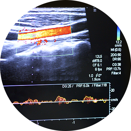
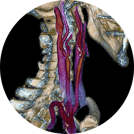
Carotid ultrasound uses sound waves to produce pictures of the carotid arteries in the neck which carry blood from the heart to the brain. A Doppler ultrasound study, a technique that evaluates blood flow through a blood vessel, is usually a part of this exam. It is most frequently used to screen patients for blockage or narrowing of the carotid arteries which may increase the risk of stroke.
Mammography is a type of low-dose X-ray of the breast. It reveals masses and micro-calcifications within and around the breast that may indicate breast problems including, but not just limited to cancer.
Experts recommend a mammography screening of the breasts at regular intervals to increase the chance of early cancer detection and treatment. The American College of Radiology recommends women aged 40 and older should have a screening mammogram every year and should continue to do so for as long as they are in good health.
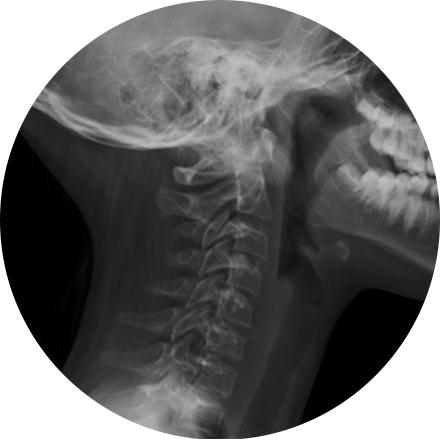
X-ray is the oldest and most economical form of medical imaging. During the procedure, radiation passes through the body onto “film” (now digitized and displayed on a computer screen). In neuroimaging, spinal X-rays are used to assess for the degree of spinal motion with flexion or extension.
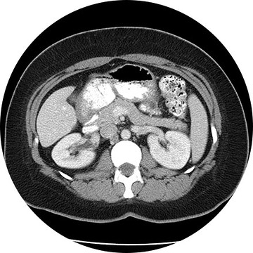
This is an accurate and noninvasive imaging procedure used to assess and evaluate certain gastrointestinal problems, such as inflammatory bowel disease (including Crohn’s Disease), infectious enteritis, lymphoma or tuberculosis. It also can be used in patients with gastrointestinal bleeding to determine if a small bowel polyp is causing the bleeding. Enterography may be performed using MRI or CT.
The biliary system is comprised of the liver, gallbladder and bile ducts, and a hepatobiliary exam, or HIDA, evaluates issues in that system. A radioactive tracer is injected (or inhaled or swallowed) and then taken up by the target organs. In this way, the clinician can see how well they function. It shows if the bile ducts are closed or leaking, or if the gallbladder is inflamed, or if the liver is abnormal. HIDA is a nuclear medicine test, which delivers information that often cannot be obtained by other procedures.
Lung cancer CT screening is one of the most accurate diagnostic tools for finding lung cancer at an early stage, when it is most treatable. CT scans of the lung are able to detect small abnormalities in the lungs that could be the beginning stages of lung cancer. These indicators are often not visible on a routine chest X-ray. Since a CT lung screening offers the best opportunity for successful treatment of lung cancer before symptoms are noticed, more physicians are opting for lung cancer screening based on risk factors (like smoking and family history), rather than symptoms.
A ventilation/perfusion lung scan (also known as a V/Q lung scan) assesses the circulation of air (ventilation) and blood (perfusion) in a patient’s lungs. The exam is useful for identifying blood clots or abnormal flow in the lungs or serious lung disorders such as chronic obstructive pulmonary disease (COPD) or a pulmonary embolism. The V/Q scan uses intravenous material (radiotracers) to show the functional health of the organs.
MRI-guided biopsy uses MRI to guide the radiologist to the exact location of the area of concern. MRI biopsy is usually used when the abnormality can be best seen on breast MRI, but not as well visualized on mammogram or ultrasound. Often lesions that are biopsied are done under the imaging modality that best demonstrates them.
Image-guided biopsy allows patients to avoid hospitalization and general anesthesia (previously necessary with traditional surgical biopsy). It is a valuable method of getting a fast, accurate, conclusive diagnosis—without unnecessary time, pain or expense to the patient.
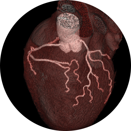
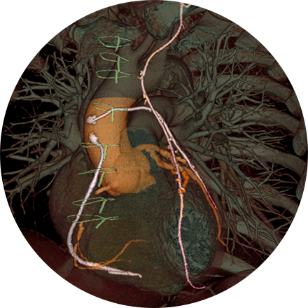
A multi-gated acquisition (MUGA) scan creates video images that show whether the lower chambers (ventricles) of the heart are pumping blood properly. MUGA uses intravenous material (radiotracers) to show how blood moves through the heart. MUGA can be used to check for pre-existing cardiac conditions prior to chemotherapy, or after treatment to assess possible side effects.
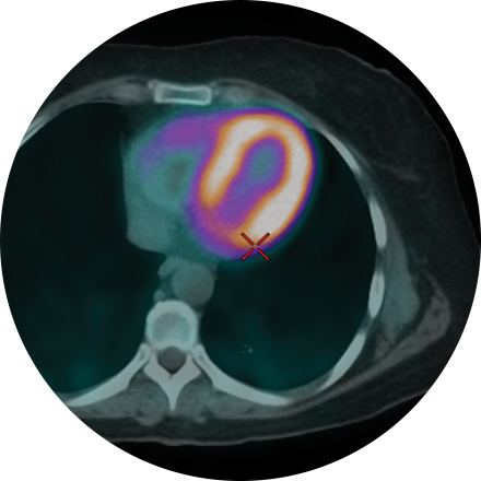
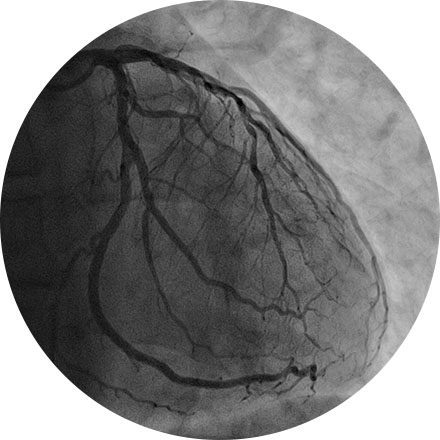
This non-invasive exam shows how well blood perfuses (flows through) your heart muscle—in other words, how well your heart is pumping. Sometimes known as a nuclear stress test, it can be performed while the patient exercises on a treadmill or, if that is inadvisable, using a medicine that simulates the effect of exercise on the heart. Myocardial perfusion is an effective way to assess narrowed arteries, the effects of a past heart attack, or the viability of further procedures, such as a stent.
One of the more frequent applications of ultrasound is in the evaluation of a fetus. Most obstetricians perform a routine diagnostic ultrasound to look for any abnormalities with either the fetus or the mother’s anatomy during early pregnancy. Depending on the time of gestation and positioning, the gender may or may not be identified. This ultrasound procedure is painless and non-invasive.
Ultrasound imaging of the pelvis uses sound waves to produce pictures of the structures and organs in the lower abdomen and pelvis. There are various types of pelvic ultrasound including abdominal, vaginal and rectal. These exams are frequently used to evaluate the reproductive and urinary systems. Ultrasound is safe, noninvasive and does not use ionizing radiation.
Ultrasound is a non-invasive procedure that uses sound waves to image the structure and movement of the body’s internal organs, as well as blood flowing through blood vessels, in real-time. The prostate gland and surrounding tissue are examined by the insertion of an ultrasound probe into the patient’s rectum. There are no harmful effects, and it gives a clearer picture of soft tissues than X-ray images.
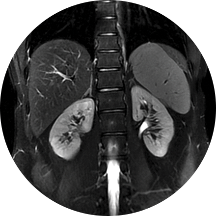
A renal ultrasound is a safe and painless test that uses sound waves to produce images of the kidneys and bladder. The kidneys are a pair of bean-shaped organs located toward the back of the abdominal cavity, just above the waist. They remove waste products from the blood and produce urine.
Ultrasound imaging of the scrotum uses sound waves to produce an image of a man’s testicles and the surrounding tissue. It is primarily used to help evaluate disorders of the testicles, and scrotum. Ultrasound does not use ionizing radiation.
CT of the sinuses uses special x-ray equipment to evaluate the hollow, air-filled spaces within the bones of the face surrounding the nasal cavity. CT scanning is painless, noninvasive and accurate. It’s also the most reliable imaging technique for determining if the sinuses are obstructed and the best imaging modality for sinusitis.
A three phase bone scan is a nuclear medicine test, it uses radiotracers that are injected (or inhaled or swallowed). The radiotracers are detected by a special camera to provide pictures to diagnose a fracture when it cannot be seen on an X-Ray. It is also used to diagnose bone infection, bone pain, osteomyelitis, as well as other bone diseases.
A thyroid scan shows, in video images, how well a patient’s thyroid is functioning, along with its structure and position. This type of study can be used to diagnose hyperthyroidism, cancer, and other abnormalities, such as lumps or inflammation, in this important organ. Like other scans that are used to determine whether an organ is functioning properly, a thyroid scan is a nuclear medicine test; that is, it uses intravenous radiotracers detected by a special camera to provide pictures that show
Thyroid ultrasound uses sound waves to produce images of the thyroid gland in the neck. This procedure is typically used to evaluate lumps (or nodules) found during a routine physical and to determine if they are the more common benign nodule or if they have features that require a biopsy. Ultrasound does not use ionizing radiation.
Transvaginal ultrasound imaging of the abdomen uses sound waves to produce pictures of the structures and organs in and around a woman's uterus. Transvaginal (TV) ultrasounds are performed very much like a gynecologic exam and involves the insertion of an ultrasound transducer into the vagina to study the lining of the uterus. These exams are frequently used to evaluate the reproductive and urinary systems. Ultrasound is safe, noninvasive and does not use ionizing radiation.