

Abdominal imaging addresses the health of all organs in your abdomen and pelvis, including the liver, gall bladder, bile ducts, spleen, pancreas, adrenal glands, kidneys, stomach, small and large intestine, aorta, inferior vena cava, male/female pelvic organs, bones and more.
Some Abdominal Imaging Procedures Include:
This is an accurate and noninvasive imaging procedure used to assess and evaluate certain gastrointestinal problems, such as inflammatory bowel disease (including Crohn’s Disease), infectious enteritis, lymphoma or tuberculosis. It also can be used in patients with gastrointestinal bleeding to determine if a small bowel polyp is causing the bleeding. Enterography may be performed using MRI or CT.
The biliary system is comprised of the liver, gallbladder and bile ducts, and a hepatobiliary exam, or HIDA, evaluates issues in that system. A radioactive tracer is injected (or inhaled or swallowed) and then taken up by the target organs. In this way, the clinician can see how well they function. It shows if the bile ducts are closed or leaking, or if the gallbladder is inflamed, or if the liver is abnormal. HIDA is a nuclear medicine test, which delivers information that often cannot be obtained by other procedures.
One of the more frequent applications of ultrasound is in the evaluation of a fetus. Most obstetricians perform a routine diagnostic ultrasound to look for any abnormalities with either the fetus or the mother’s anatomy during early pregnancy. Depending on the time of gestation and positioning, the gender may or may not be identified. This ultrasound procedure is painless and non-invasive.
Ultrasound imaging of the pelvis uses sound waves to produce pictures of the structures and organs in the lower abdomen and pelvis. There are various types of pelvic ultrasound including abdominal, vaginal and rectal. These exams are frequently used to evaluate the reproductive and urinary systems. Ultrasound is safe, noninvasive and does not use ionizing radiation.
Ultrasound is a non-invasive procedure that uses sound waves to image the structure and movement of the body’s internal organs, as well as blood flowing through blood vessels, in real-time. The prostate gland and surrounding tissue are examined by the insertion of an ultrasound probe into the patient’s rectum. There are no harmful effects, and it gives a clearer picture of soft tissues than X-ray images.
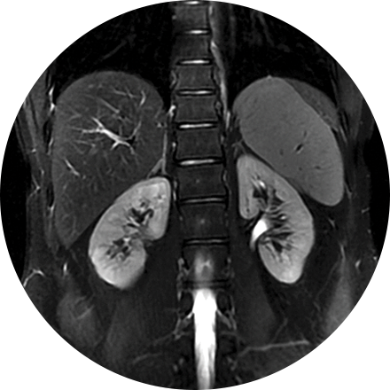
A renal ultrasound is a safe and painless test that uses sound waves to produce images of the kidneys and bladder. The kidneys are a pair of bean-shaped organs located toward the back of the abdominal cavity, just above the waist. They remove waste products from the blood and produce urine.
A three phase bone scan is a nuclear medicine test, it uses radiotracers that are injected (or inhaled or swallowed). The radiotracers are detected by a special camera to provide pictures to diagnose a fracture when it cannot be seen on an X-Ray. It is also used to diagnose bone infection, bone pain, osteomyelitis, as well as other bone diseases.