


Case Study – OHVIRA Syndrome
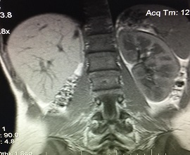
Imaging Findings
MRI Findings were crucial in determining the correct diagnosis in this patient with a complex congenital anomaly consisting of uterine didelphys, obstructed right hemivagina (atresia) with hematocolpus, ipsilateral agenesis of right kidney, and non-obstructed left hemivagina.
FINAL DIAGNOSIS – OHVIRA SYNDROME (Obstructed Hemivagina with Ipsilateral Renal Anomaly)
Discussion
OHVIRA syndrome (previously known as Herlyn-Werner-Wunderlich syndrome) was first reported in 1950 [1] and is a rare congenital anomaly of the Müllerian (paramesonephric) ducts and Wolffian (mesonephric) structures consisting of the following triad: obstructed hemivagina, ipsilateral renal anomaly, and uterine didelphys. Typically, patients present during puberty with nonspecific/variable symptoms in addition to pelvic pain shortly following menarche, which can cause a delay in the diagnosis. Symptoms include worsening dysmenorrhea, pelvic pain, and a pelvic mass and some patients may present with urgency, frequency, or vaginal discharge [2].
Müllerian duct anomalies (MDAs) have an incidence of 2–3% and OHVIRA syndrome makes up 0.16–10% of these MDAs [2]. These are due to non-development (agenesis or hypoplasia), defective vertical or lateral fusion (lateral defects tend to be the most common), or resorption failure of the Müllerian ducts. Hypoplasia and agenesis of the uterus and proximal vagina account for 5%-10% of MDAs, whereas didelphys uterus accounts for approximately 11% of MDAs [3]. In addition, anomalies of the ipsilateral renal tract coincide with MDAs in up to 30% of cases [3].
Although it contains defects in differing embryonic lineages, OHVIRA syndrome possesses paramesonephric and mesonephric defects (uterine and renal defects), which are due to embryologic arrest in the 8th week of gestation [4]. Other than this known embryologic arrest, the exact cause of the arrest is not known. However, if one of the Wolffian ducts is absent, the Wolffian structures (kidney and ureter) on the ipsilateral side will fail to fuse at midline, which may be a complete or incomplete fusion failure. In a complete failed fusion, a uterus didelphys forms. The Müllerian duct, on the side without the Wolffian duct, displaces itself laterally and is not able to come into contact with the urogenital sinus in the center, which results in a blind sac, imperforate, or obstructed vagina. A complete or partial vaginal septum is present in 75% of women with didelphys uterus [3]. The distal part of the vagina, however, originates from the urogenital sinus and thus is not affected. Each ureteric bud develops from the Wolffian duct and is essentially responsible for the formation the kidney. Occasionally an ectopically obstructed ureter inserts into the obstructed hemivagina on the side of the congenitally absent ipsilateral kidney. The ureteric bud grows dorsocranially into the metanephric blastema and induces the differentiation of the metanephric nephrons. If the ureteric bud is not able to either form or come into contact with the metanephric blastema, the kidney on that side will fail to develop [2], which thus describes the renal agenesis often found with OHVIRA syndrome.
Awareness of these syndrome variants is important for proper diagnosis and therapeutic management decisions. For instance, the treatment of a patient diagnosed with double uterus and obstructed hemivagina is generally limited to surgical correction with hysteroscopic excision of the obstructive septum located between the obstructed hemivagina and normal hemivaginas, whereas other possible variants such as unilateral cervical atresia require a more complex surgical correction.
Bibliography:
-
A Case of Uterus Didelphys with Unilateral Gynatresia. Br Med J. 1950;1(4657):820
-
Cox D, Ching BH. Herlyn-Werner-Wunderlich syndrome: a rare presentation with pyocolpos. J Radiol Case Rep. 2012;6(3):9-15
-
Del vescovo R, Battisti S, Di paola V, et al. Herlyn-Werner-Wunderlich syndrome: MRI findings, radiological guide (two cases and literature review), and differential diagnosis. BMC Med Imaging. 2012;12(1):4
-
Amin El-Gohary, Mohamed. “Uterus Didelphys with Obstructed Hemivagina and Ipsilateral Renal Anomaly (OHVIRA Syndrome): A Case Report.” ScienceDirect. Journal of Pediatric Surgery Case Reports, 1 Sept. 2014. Web. 22 Dec. 2015.
Authors’ information
-
Joshua Ian Gottlieb; NYIT College of Osteopathic Medicine; Northern Boulevard; Old Westbury, NY, 11568; joshgottlieb@sbcglobal.net.
-
Roy Gottlieb, D.O., FSCCT; Medical Director, Rolling Oaks Radiology; 415 Rolling Oaks Drive, Suite 125, Thousand Oaks, CA 91361; (818) 398-4928 cell; (805) 778-1513 work; roygottlieb@sbcglobal.net ; corresponding author.


Add new comment