


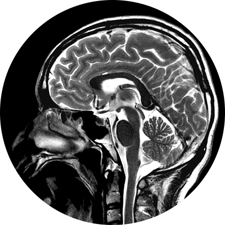
Our board-certified neuroradiologists investigate pathologies and injuries of the head, neck, and spine using the most advanced imaging technologies available. Using mainly MRI and CT, neuroradiologists diagnose abnormalities of the central and peripheral nervous system.
Some Brain & Spine Imaging Procedures Include:
Carotid ultrasound uses sound waves to produce pictures of the carotid arteries in the neck which carry blood from the heart to the brain. A Doppler ultrasound study, a technique that evaluates blood flow through a blood vessel, is usually a part of this exam. It is most frequently used to screen patients for blockage or narrowing of the carotid arteries which may increase the risk of stroke.
A FDG brain PET/CT is used to image the metabolic function of the brain. Positron emission tomography (PET) uses small amounts of radioactive materials called radiotracers, a special camera, and a computer to help evaluate the brain. It is most commonly used to calculate the degree and pattern of brain volume loss, which allows the neuroradiologist to assess for different types of neurodementia syndromes. It can also help differentiate recurrent brain tumor vs. radiation change after a patient has had brain surgery. Additionally, it can help identify the seizure focus in patients with epilepsy.
Myelography is an exam in which contrast material is injected into your spinal column and then that contrast and spinal anatomy is imaged with CT technique. This allows the neuroradiologists to evaluate areas of nerve root impingement, canal narrowing, or disc protrusions. It is typically used when a patient is not a candidate for MRI.
NeuroQuant (NQ) is an artificial intelligence (AI) tool that calculates the volume of different substructures of the brain and compares those to a large normative age- and gender-matched database to determine whether the degree of brain volume loss is statistically significant for patient age. This can be used to improve the early detection of Alzheimer's Disease (AD) or other neurodementia syndromes. NeuroQuant-MS is used to calculate the volume, number, and location of plaques in patients with multiple sclerosis. The software highlights new, enlarging, or shrinking plaques. This allows for accurate tracking of disease status over time. NQ can also be used to detect the location of a seizure focus in patients with epilepsy. It is also used in brain trauma patients or to assess brain development. Neuro-Quant has both MRI and CT applications. Other NeuroQuant-based AI tools in development include volumetric quantification and characterization of brain tumors.
CT of the sinuses uses special x-ray equipment to evaluate the hollow, air-filled spaces within the bones of the face surrounding the nasal cavity. CT scanning is painless, noninvasive and accurate. It’s also the most reliable imaging technique for determining if the sinuses are obstructed and the best imaging modality for sinusitis.
A thyroid scan shows, in video images, how well a patient’s thyroid is functioning, along with its structure and position. This type of study can be used to diagnose hyperthyroidism, cancer, and other abnormalities, such as lumps or inflammation, in this important organ. Like other scans that are used to determine whether an organ is functioning properly, a thyroid scan is a nuclear medicine test; that is, it uses intravenous radiotracers detected by a special camera to provide pictures that show
Thyroid ultrasound uses sound waves to produce images of the thyroid gland in the neck. This procedure is typically used to evaluate lumps (or nodules) found during a routine physical and to determine if they are the more common benign nodule or if they have features that require a biopsy. Ultrasound does not use ionizing radiation.

