
Imaging Modalities Help Researchers See into the Past
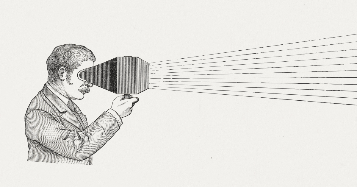
At RadNet centers, we use an array of imaging modalities to help patients get the healthcare answers they need. Services such as PET/CT, MRI, ultrasound and digital X-ray give radiologists a better view inside our bodies - at our brains, hearts, muscles, spines, organs and bones – but did you know that these imaging techniques have also been used in recent years by experts such as scientists, archaeologists, historians and researchers to help them see into the past?
Imaging technology has advanced to such specificity that MRI can actually provide the highest level of soft tissue detail, or a PET exam of the brain can help diagnose Alzheimer’s disease up to 20 years before a patient becomes symptomatic, plus many other incredible breakthroughs. In the same way these developments have helped propel healthcare for people, they have also helped scientists see things never before possible. Let’s take a look at some of these amazing developments.
Dental X-Rays Used to Read Sealed 17th Century Letter
An article published in Nature Communications describes how researchers were able to virtually unfold and read a sealed letter from the Renaissance era in Europe. These researchers, from the Queen Mary University of London’s dental research labs, developed a scanner to scan a batch of unopened letters found in a 17th century postal trunk that had never been delivered.
The 300 year-old letters had originally been sealed using “letter-locking,” a common bygone way of folding up a sheet of paper into its own envelope. Until now, people who found these old letters had to cut them open to read them, which lead to damaging the contents of the letters themselves.
Through this new x-ray process, researchers opened one of these letters and found that it was dated July 31, 1697. In the letter, Jacques Sennacques asked his cousin, Pierre Le Pers, a French merchant in the Hague, for a certified copy of a death notice for Daniel Le Pers.
Ongoing CT Mummy Project
Originally launched in 2005, this project studies human remains from Egyptian pyramids with the goal of shedding light on both the royal individuals examined and on the civilization in general, including its science, mummification methods, and the prevalence and treatment of disease.
According to an article in Aunt Minnie, researchers have been able to pinpoint cause of death for many ancient kings using CT exams. For instance, they were able to determine that Ramses III was killed by multiple assailants, due to evidence of several wounds and injuries that were only visible on the CT exam.
The mummies of ancient Egypt also offer a wealth of information regarding the history of disease. Researchers discovered that ancient Egyptian royal families suffered from a degenerative spinal disease (DISH), rather than from an inflammatory arthritis, which had been previously diagnosed from plain film x-rays. This research may help illuminate how diseases develop and the ways modern people could be affected.
CT also helped identify hidden objects and amulets that were secreted inside the mummified bodies of royalty or inside of their wrappings. One CT exam showed that Thuya, great-grandmother of Tutankhamun, was wearing golden sandals, hidden beneath her wrappings.
X-Ray Finds the Source of the Middle Ages “Dark Clouds”
The year was 536. According to historical accounts, a mysterious fog plunged Europe, the Middle East and parts of Asia into darkness, day and night. Temperatures plummeted, snow fell in the summer, crops died, people starved. Then, the bubonic plague spread, wiping out one-third to one-half of the population. Historians have long known that the middle of the sixth century was a dark hour in what used to be called the Dark Ages, but the source of dark clouds has long been a puzzle. According to an article in Science Magazine, a team from the Climate Change Institute of The University of Main (UM) in Orono has finally found an answer.
The research team reported that a cataclysmic volcanic eruption in Iceland spewed ash across the Northern hemisphere in early 536. Two other massive eruptions followed, in 540 and 547.
The interdisciplinary team drilled an ice core in an glacier in the Swiss Alps. The 72-meter-long core entombs more than 2000 years of fallout from volcanoes, Saharan dust storms and human activities. The team deciphered the core using a new ultra-high-resolution method, in which a laser carves slivers of ice which each represent just a few days or weeks of snowfall. Each of the samples is analyzed for about a dozen elements using sophisticated x-ray equipment. The approach enabled the team to pinpoint storms, volcanic eruptions and lead pollution down to the month or even less, going back 2000 years.
When the ice from the spring of 536 was analyzed, researchers found two microscopic particles of volcanic glass. These shards were bombarded with x-rays to determine their chemical fingerprint, which closely matched glass particles found earlier in other locations, which in turn, resembled volcanic rocks from Iceland.
Ultimately, researchers have found through x-ray that the frozen ice core provides a detailed log of natural disasters and human pollution.
In addition to these fascinating discoveries, screening modalities have also been used to help researchers learn more about dinosaurs, uncover lost inscriptions on ancient artifacts, unearth ancient rock paintings – and many other amazing findings that could not have been possible without the incredible advancements that have been made in radiology and imaging technology.