
Breast cancer is the second most common cancer in women (after lung cancer). Early detection of breast cancer vastly increases survival rates. Scheduling an annual mammogram after the age of 40 (or earlier, depending on your breast cancer risk factors) is one of the most important things women can do to ensure to take control of their health.
Some Breast Imaging Procedures Include:
Your provider may request a breast biopsy if a lump or abnormality is found during screening. A biopsy is a very accurate, minimally invasive method of locating and removing tissue for further investigation. Image-guided biopsy allows patients to avoid hospitalization and general anesthesia (previously necessary with traditional surgical biopsy). It is a valuable method of getting a fast, accurate, conclusive diagnosis—without unnecessary time, pain or expense to the patient.
Depending on your medical needs, we provide different diagnostic biopsy procedures:
-
Stereotactic biopsy uses mammography to precisely guide clinicians to a lump or abnormality that can’t be felt or seen on ultrasound. Clinicians see a 3D picture of the lump’s exact location.
-
MRI-guided biopsy uses MRI to guide the radiologist to the exact location of the lump. MRI biopsy is usually used when the lump can be seen on breast MRI, but cannot be readily seen on mammogram or ultrasound.
What happens during a biopsy procedure?
While the biopsy is performed, you will either remain seated in a comfortable, upright position or you will lie face down on a special table that allows your breast to be placed in an opening. The radiologist performs a core needle or vacuum-assisted biopsy (see below) while your breast is somewhat compressed in the mammographic biopsy system. You can return home in 30 minutes.
-
Vacuum-assisted biopsy devices are used for stereotactic and MRI guided biopsy and selected ultrasound guided biopsies. The device is a special probe that applies suction and allows retrieval of more tissue. Breast tissue is drawn into the sampling chamber of the probe with the vacuum and then cut. Several pieces of tissue are always obtained during biopsies, regardless of the type of needle used.
-
In a core needle biopsy, the radiologist locates the lump or abnormality that can be seen on a mammogram, sonogram or MRI. A hollow core needle is then placed inside the abnormality. The needle will then withdraw a small amount of tissue that will be sent to a lab for analysis. Prior to the procedure, you will be given some local anesthetic similar to the anesthetic used for dental procedures to numb the area. You may feel some pressure and mild discomfort but most patients do not feel pain. The doctor will insert the needle several times to get adequate tissue samples. After the procedure, you can resume normal activities immediately. You may experience some slight bruising but should not have a scar. It may take several days to one week to obtain results from the lab.
Some women — because of their family history, a genetic tendency or certain other factors — should be screened with MRI in addition to mammograms. The number of women who fall into this category is small: less than 2 percent of all the women in the United States. Talk with your doctor about your history and whether you should have additional tests.
Breast MRI helps to detect small lesions sometimes missed by mammography — without using radiation or compressing the breasts. MRI uses powerful magnets and radio waves to take very clear pictures of soft tissues, so it can be extremely useful in breast imaging.
Ultrasound uses sound waves instead of X-rays to exam breast tissue. A transducer is passed over the breast. The transmitted sound waves are translated into a picture on a monitor. It does not cause discomfort and, because it does not use radiation, it carries very little risk.
Ultrasound is useful for women with dense breasts or for evaluating suspicious areas seen by mammography or felt during a breast exam. It can also find breast lesions that are close to the chest, where mammography is less useful. Breast ultrasound can distinguish between cysts, which are fluid-filled, versus other types of solid breast masses.
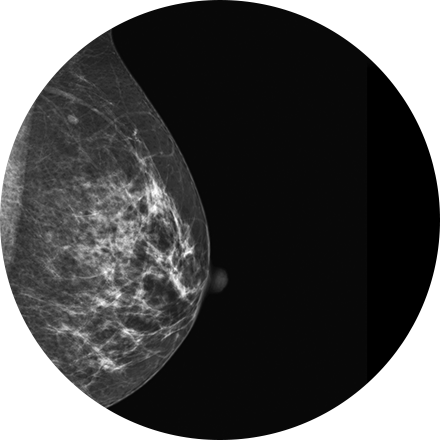
Mammography is a type of low-dose X-ray of the breast. It reveals masses and micro-calcifications within and around the breast that may indicate breast problems including, but not just limited to cancer.
Experts recommend a mammography screening of the breasts at regular intervals to increase the chance of early cancer detection and treatment. The American College of Radiology recommends women aged 40 and older should have a screening mammogram every year and should continue to do so for as long as they are in good health.