
Interventional radiology is the practice of performing minimally invasive procedures to diagnose and treat diseases and disorders. We provide a number of these services, which can sometimes help patients avoid more complicated surgical interventions. Patients have less pain and shorter recovery periods.
Common Interventional Radiology Procedures
Our interventional radiologists perform such a wide range of procedures, some of which treat blood vessels (vascular procedures), that it is difficult to list them all. Some common interventional radiology procedures are:
This is the use of CT, ultrasound, MRI or fluoroscopy to precisely locate and evaluate masses or suspicious areas throughout the body. Radiologists use tiny instruments to take small tissue samples that are evaluated for cancer. During a surgical biopsy, radiologists help surgeons locate the mass using a guide wire.
Your provider may request a breast biopsy if a lump or abnormality is found during screening. A biopsy is a very accurate, minimally invasive method of locating and removing tissue for further investigation. Depending on your medical needs, we provide different diagnostic biopsy procedures:
-
Stereotactic biopsy uses mammography to precisely guide clinicians to a lump or abnormality that can’t be felt or seen on ultrasound. Clinicians see a 3D picture of the lump’s exact location.
-
MRI-guided biopsy uses MRI to guide the radiologist to the exact location of the lump. MRI biopsy is usually used when the lump can be seen on breast MRI, but cannot be readily seen on mammogram or ultrasound.
What happens during the procedure?
While the biopsy is performed, you will either remain seated in a comfortable, upright position or you will lie face down on a special table that allows your breast to be placed in an opening. The radiologist performs a core needle or vacuum-assisted biopsy (see below) while your breast is somewhat compressed in the mammographic biopsy system. You can return home in 30 minutes.
-
Vacuum-assisted biopsy devices are used for stereotactic and MRI guided biopsy and selected ultrasound guided biopsies. The device is a special probe that applies suction and allows retrieval of more tissue. Breast tissue is drawn into the sampling chamber of the probe with the vacuum and then cut. Several pieces of tissue are always obtained during biopsies, regardless of the type of needle used.
-
In a core needle biopsy, the radiologist locates the lump or abnormality that can be seen on a mammogram, sonogram or MRI. A hollow core needle is then placed inside the abnormality. The needle will then withdraw a small amount of tissue that will be sent to a lab for analysis. Prior to the procedure, you will be given some local anesthetic similar to the anesthetic used for dental procedures to numb the area. You may feel some pressure and mild discomfort but most patients do not feel pain. The doctor will insert the needle several times to get adequate tissue samples.
Vertebroplasty and kyphoplasty are procedures that treat spinal fractures or compressed / collapsed vertebrae, often performed by a neuroradiologist. Vertebroplasty is the injection of a cement-like material into the bone to make it more stable. In kyphoplasty, the doctor first creates space by inflating a balloon-like device in the bone. The space is then filled with the cement material.
Uterine Fibroid Embolization (UFE) decreases the pain and bleeding of uterine fibroids (benign tumors from the wall of the uterus) by cutting off their blood supply.
Pelvic venography helps diagnose women whose pelvic pain is caused by swelling of pelvic veins (pelvic congestion syndrome). To treat the condition, the radiologist inserts a thin catheter, about the size of a strand of spaghetti, into the femoral vein in the groin and guides it to the affected vein using X-ray guidance. To seal the faulty, enlarged vein and relieve painful pressure, an interventional radiologist inserts tiny coils often with a sclerosing agent (the same type of material used to treat varicose veins) to close the vein.
This procedure allows doctors and nurses to easily access the venous system to repeatedly draw blood or to inject substances into the body. This procedure is useful when you need ongoing chemotherapy, antibiotic therapy, parenteral nutrition or kidney dialysis. In these procedures, the radiologist uses ultrasound to guide the placement of a thin catheter or other device into your chest, neck, or arm. Some common vein access procedures include:
-
Kidney dialysis access management—Central venous access is increasingly used in kidney dialysis.
-
Mediports (also called port-a-caths)—Devices are usually implanted in the upper chest to act as an I.V. into the bloodstream; they are used to deliver chemotherapy, medications, fluids, or to withdraw blood.
-
PICCs (peripherally inserted central catheters)—Catheters inserted into the arm vein are threaded carefully to the superior vena cava, the large vein that connects to the heart. PICCs are used for long-term intravenous antibiotics, chemotherapy, etc. and to prevent repeat needle sticks.
CT or other imaging tools are used to guide the insertion of needles or tubes to drain abscesses.
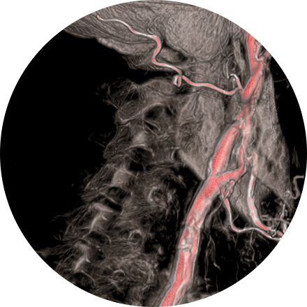
Angiography is a common interventional radiology procedure that can help doctors diagnose blockages, bleeding or other disorders in blood vessels throughout the body. During an angiogram, the radiologist inserts a tiny catheter into a blood vessel using a minute puncture in your skin, then injects a dye to make the blood vessels visible during a special type of X-ray called fluoroscopy.
This procedure is used to stop unwanted bleeding, as with a tumor, injury, fistula, or aneurysm. Medications or synthetic materials (called embolic agents) are placed through a catheter into a blood vessel to prevent blood flow to the area.
This is minimally invasive treatment of varicose veins in one or more outpatient procedures.
Needle electrodes are inserted through the skin and into the site of a tumor in the kidney, liver, prostate or lungs. The radio waves emitted by the probe create heat in the tissue, and the heat kills cancerous cells in a small area around the probe.
Interventional procedures often provide invaluable assistance to your urologist or other physician for problems of the genitals and urinary tract. A few of the common procedures we perform include:
-
Nephrostomy – Placement of a tube into the kidney to allow urine to drain directly out of the body.
-
Kidney stone treatment – Manipulation and management of kidney stones with minimally invasive techniques.
-
Biliary procedures – Viewing and/or treating problems of the bile ducts, liver and gallbladder, including bile stones. These procedures include percutaneous cholangiogram (a contrast x-ray to find obstructions in the bile ducts), biliary stone removal (when they cannot be removed surgically) and biliary stent placement (for temporary relief from bile stone obstructions).
Some Interventional Radiology Imaging Procedures Include:
An angiogram is an X-ray exam of the arteries and veins to diagnose blockages and other blood vessel problems. It can reveal the integrity of the cardiovascular system in specific areas throughout the body. Combined with the use of intravenous contrast medium injected via a catheter, an angiogram identifies areas of blockage or damaged vessels within the circulatory system. CT and MRI may also be used to gain additional images of the arteries.
Biopsies (removal of tissue for investigation) may be performed with the guidance of CT, MR, ultrasound or X-ray images. If a lesion is discovered, a bone biopsy is performed through the skin to determine whether cancer or infection is present.
Your provider may request a breast biopsy if a lump or abnormality is found during screening. A biopsy is a very accurate, minimally invasive method of locating and removing tissue for further investigation. Image-guided biopsy allows patients to avoid hospitalization and general anesthesia (previously necessary with traditional surgical biopsy). It is a valuable method of getting a fast, accurate, conclusive diagnosis—without unnecessary time, pain or expense to the patient.
Depending on your medical needs, we provide different diagnostic biopsy procedures:
-
Stereotactic biopsy uses mammography to precisely guide clinicians to a lump or abnormality that can’t be felt or seen on ultrasound. Clinicians see a 3D picture of the lump’s exact location.
-
MRI-guided biopsy uses MRI to guide the radiologist to the exact location of the lump. MRI biopsy is usually used when the lump can be seen on breast MRI, but cannot be readily seen on mammogram or ultrasound.
What happens during a biopsy procedure?
While the biopsy is performed, you will either remain seated in a comfortable, upright position or you will lie face down on a special table that allows your breast to be placed in an opening. The radiologist performs a core needle or vacuum-assisted biopsy (see below) while your breast is somewhat compressed in the mammographic biopsy system. You can return home in 30 minutes.
-
Vacuum-assisted biopsy devices are used for stereotactic and MRI guided biopsy and selected ultrasound guided biopsies. The device is a special probe that applies suction and allows retrieval of more tissue. Breast tissue is drawn into the sampling chamber of the probe with the vacuum and then cut. Several pieces of tissue are always obtained during biopsies, regardless of the type of needle used.
-
In a core needle biopsy, the radiologist locates the lump or abnormality that can be seen on a mammogram, sonogram or MRI. A hollow core needle is then placed inside the abnormality. The needle will then withdraw a small amount of tissue that will be sent to a lab for analysis. Prior to the procedure, you will be given some local anesthetic similar to the anesthetic used for dental procedures to numb the area. You may feel some pressure and mild discomfort but most patients do not feel pain. The doctor will insert the needle several times to get adequate tissue samples. After the procedure, you can resume normal activities immediately. You may experience some slight bruising but should not have a scar. It may take several days to one week to obtain results from the lab.
This procedure allows doctors and nurses to easily access the venous system to repeatedly draw blood or to inject substances into the body. This procedure is useful when you need ongoing chemotherapy, antibiotic therapy, parenteral nutrition or kidney dialysis. In these procedures, the radiologist uses ultrasound to guide the placement of a thin catheter or other device into your chest, neck, or arm. Some common vein access procedures include:
-
Kidney dialysis access management – Central venous access is increasingly used in kidney dialysis.
-
Mediports (also called port-a-caths) – Devices are usually implanted in the upper chest to act as an I.V. into the bloodstream; they are used to deliver chemotherapy, medications, fluids, or to withdraw blood. PICCs (peripherally inserted central catheters) – Catheters inserted into the arm vein are threaded carefully to the superior vena cava, the large vein that connects to the heart.
-
PICCs are used for long-term intravenous antibiotics, chemotherapy, etc. and to prevent repeat needle sticks.
Embolization is used to stop unwanted bleeding, as with a wound or aneurysm (ruptured or unruptured). Medications or synthetic materials (embolic agents) are placed through a catheter into a blood vessel to prevent blood flow to the affected area.
Interventional procedures often provide invaluable assistance to your urologist or other physician for problems of the genitals and urinary tract. A few of the common procedures we perform include:
-
Nephrostomy – Placement of a tube into the kidney to allow urine to drain directly out of the body.
-
Kidney stone treatment – Manipulation and management of kidney stones with minimally invasive techniques.
-
Biliary procedures – Viewing and/or treating problems of the bile ducts, liver and gallbladder, including bile stones. These procedures include percutaneous cholangiogram (a contrast x-ray to find obstructions in the bile ducts), biliary stone removal (when they cannot be removed surgically) and biliary stent placement (for temporary relief from bile stone obstructions).
Pelvic venography helps diagnose women whose pelvic pain is caused by swelling of pelvic veins (pelvic congestion syndrome). To treat the condition, the radiologist inserts a thin catheter, about the size of a strand of spaghetti, into the femoral vein in the groin and guides it to the affected vein using X-ray guidance. To seal the faulty, enlarged vein and relieve painful pressure, an interventional radiologist inserts tiny coils often with a sclerosing agent (the same type of material used to treat varicose veins) to close the vein.
Needle electrodes are inserted through the skin and into the site of a tumor in the kidney, liver, prostate or lungs. The radio waves emitted by the probe create heat in the tissue, and the heat kills cancerous cells in a small area around the probe.
CT or other imaging tools are used to guide the insertion of needles or tubes to drain abscesses.
Uterine Fibroid Embolization (UFE) decreases the pain and bleeding of uterine fibroids (benign tumors from the wall of the uterus) by cutting off their blood supply.
This is minimally invasive treatment of varicose veins in one or more outpatient procedures.
Vertebroplasty and kyphoplasty are procedures that treat spinal fractures or compressed / collapsed vertebrae, often performed by a neuroradiologist. Vertebroplasty is the injection of a cement-like material into the bone to make it more stable. In kyphoplasty, the doctor first creates space by inflating a balloon-like device in the bone. The space is then filled with the cement material.