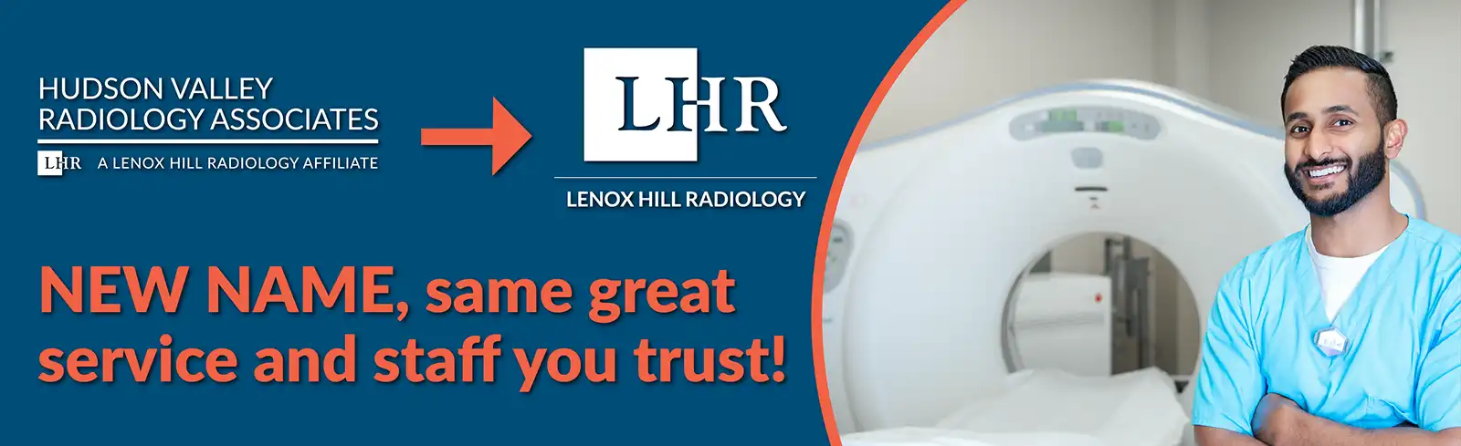
button-radnet
FIND AN IMAGING CENTER
×
 

The Hudson Valley Radiology Associates (HVRA) Advanced Cardiac Imaging Program is Hudson Valley region’s most robust, non-invasive, outpatient, cardiac imaging program.

Our Cardiac Radiologists

Cost Advantage

From HVRA’s wide-range of advanced, Low-Dose Cardiac CT units and dozens of upgraded Wide-Open Cardiac MRIs, to our highly-trained clinical and support staff, our Advanced Cardiac Imaging Program offers cardiology patients only the very best in diagnosis.

As an independent medical imaging provider operating outside of hospital affiliations, Hudson Valley Radiology Associates offers affordable, high-quality, non-invasive cardiac imaging at a cost that is up to 50% lower than that of hospitals.

Safety & Comfort is our Top Priority

Early detection can lead to additional treatment options and a more favorable outcome. Our cardiac imaging program can provide a comprehensive evaluation of the heart, including its structure, function, blood flow, and tissue perfusion.

Our Cardiac CT exams are performed with low-dose protocols on the most-advanced units. Advanced imaging can help a cardiologist tailor a treatment plan to each patient's needs, based on their individual cardiac anatomy and function.

Accurate Diagnosis

Advanced cardiac imaging techniques, such as Coronary Computed Tomography Angiography (Coronary CTA), provide several advantages, including a more accurate diagnosis.

The HVRA's Advanced Cardiac Imaging Program employs the most advanced cardiac imaging techniques and tools available in order to provide the highest-quality images of the heart structures and morphology, as well as the newest platforms and tools that help demonstrate the physiologic significance of coronary artery disease, if present. Our robust data allows our experienced cardiac radiologist to make the most accurate diagnosis of various heart conditions.

Scheduling

Cardiac Scheduling

Phone: (845) 770-6160

Email: hvraphysicianservices@radnet.com

CT Cardiac Calcium Scoring

CT Cardiac Calcium Scoring

What is CT Cardiac Calcium Scoring?

One of HVRA’s most accessible and valuable cardiac offerings is our low-cost, Low-Dose CT Cardiac Calcium Scoring exam.

Covered by most insurances, CT Cardiac Calcium Scoring is a fast, non-invasive, screening exam. It is a first-line heart test that uses Computerized Tomography (CT) to detect calcium deposits in the coronary arteries of the heart. A higher coronary calcium-score suggests you have a greater chance of clinically significant narrowing of the coronary arteries and potentially a higher risk of future heart attack.

It is important to note that a CT Cardiac Calcium Scoring exam is not diagnostic in itself. It is a screening exam and is typically used as a first line of detection and in combination with clinical evaluation by a cardiologist and other diagnostic tests, such as Coronary CTA (CCTA), to identify and evaluate the patient's risk of developing heart disease.

Benefits:

  • Non-invasive

  • 1 minute study

  • Very low radiation

  • Used for first-line screening of the coronary artery for calcified plaques

Coronary CTA (CCTA)

Coronary CTA (CCTA)

What is CCTA?

If calcified plaque is detected in the coronary arteries, a referring provider may recommend a patient for a non-invasive diagnostic CT scan of the heart, called a Coronary CT Angiography (CCTA) that provides highly detailed 3D images of the coronary arteries and anatomic data about the structure (the lumen) of the coronary arteries. This exam is typically used to detect narrowed or blocked coronary arteries, which may cause chest pain or a heart attack.

Benefits:

  • Non-invasive

  • No radiation is left in your body after the scan is finished

  • Provides very detailed images of many types of tissue

  • Fast and simple

  • Cost-effective for a wide range of medical problems

  • Less sensitive to patient movement than MRI

  • Implanted medical device of any kind will not prevent you from having a CT scan

For patients who have:

  • Suspected abnormal anatomy of the coronary arteries

  • Low or intermediate risk for coronary artery disease, including patients who have chest pain and normal, non-diagnostic or unclear lab and ECG results

  • Low to intermediate risk atypical chest pain in the emergency department

  • Non-acute chest pain

  • New or worsening symptoms with a previous normal stress test result

  • Unclear or inconclusive stress test results

  • New onset heart failure with reduced heart function and low or medium risk for coronary artery disease

  • Intermediate risk of coronary artery disease before non-coronary cardiac surgery

  • Coronary artery bypass grafts


American Heart Association and the American College of Cardiology Risk Calculator

CCTA with FFR CT (Fractional Flow Reserve)

CCTA with FFR CT (Fractional Flow Reserve)

How CCTA with FFR CT works

Coronary CTA with Fractional Flow Reserve CT (CCTA w/ FFR CT)
 

CT with Fractional Flow Reserve (FFR CT) is a physiologic simulation technique that models coronary flow from routine Coronary CT Angiography (CTA). At Hudson Valley Radiology Associates, FFR CT is always interpreted in correlation with clinical and anatomic coronary CTA findings.


FFR CT increases the specificity in the detection and evaluation of coronary artery disease, and decreases the prevalence of non-obstructive disease in invasive coronary angiography (ICA), which helps with decisions regarding revascularization vs. pharmacologic or other forms of treatment.

How can CCTA with FFR CT help:

  • Without additional patient tests, the Fractional Flow Reserve Analysis quickly and non-invasively delivers functional information (FFR CT values) about each blockage

  • Completing the picture for each patient leads to better clinical decision-making and improved patient outcomes

  • Recognized in ACC/AHA Chest Pain Guidelines to help guide treatment for patients with CAD

Coronary CTA with Fractional Flow Reserve CT (CCTA w/ FFR CT)
 

Demonstrates:

  • Diagnostic confidence that delivers increased per-vessel diagnostic performance relative to other non-invasive cardiac tests

  • Visualize disease other non-invasive cardiac tests may miss

  • Enables physicians to confidently identify patients who can be treated optimally with pharmacologic therapies alone

  • Enables providers to present patients a compelling visual understanding of their disease state and impact it has on their heart

CCTA with AI Plaque Analysis

CCTA with AI Plaque Analysis

How CCTA with AI Plaque Analysis works

If your cardiologist recommends a CCTA with AI plaque analysis, a standard CCTA can be processed through machine learning software to analyze atherosclerosis (plaque) and stenosis for an additional cost (not covered by insurance). The AI Plaque Analysis generates a 3D model of the coronary arteries, identifies their lumen and vessel walls, locates and quantifies stenoses, and identifies, quantifies and categorizes plaque. This additional process helps clinicians precisely identify and define atherosclerosis earlier.

CCTA with AI plaque analyses

How can CCTA with AI plaque analysis help:

  • Non-invasive tests for patients that may help identify issues before symptoms occur

  • Cardiac providers may improve evaluation and individualized approach to their patient's cardiovascular care

  • Employers can improve employee health outcomes and reduce potential healthcare costs


Cardiac Radiologists

Roger Frey, M.D.

Roger Frey
M.D.

Click Here to Learn More


You Can't Beat a Healthy Heart!