
What is PET/MRI?
A PET/MRI scan is a two-in-one test that combines images from a positron emission tomography scan and a magnetic resonance imaging (MRI) scan. This hybrid technology utilizes both PET and MRI to produce some of the most highly detailed pictures of the inside of your body currently available. Doctors use those pictures to diagnose medical conditions and plan treatment.
When is PET/MRI used?
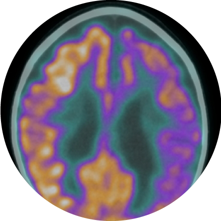
PET/MRI is a recent addition to imaging in an out-patient setting, it is particularly useful in diagnosing oncologic and neurological conditions.
Other situations where PET/MRI might be useful include:
-
The effect of certain medications on the heart and brain.
-
Abnormal tissue or tumors.
-
Early recognition of dementia.
-
Shifts in blood distribution.
-
Areas affected by a stroke or blood clot.
What happens during a PET/MRI procedure?
MRI scans use a strong magnetic field to produce detailed images of internal structures in the body. They can also provide information about how well these structures are functioning. PET scans use tracers to highlight abnormalities that may indicate disease. These combined create a 3D image of the body. The entire procedure usually lasts between two to three hours.
What are the benefits and risks?
PET/MRI can provide a more accurate diagnosis and therefore better treatment options. By combining the two modalities, it also makes it easier to get the scans done during a single appointment instead of having to book multiple. The risks involved with the procedure are minimal as there is no radiation involved.