

Imaging procedures for the brain can discern both the function and the structure of the nervous system. Also known as neuroimaging, these types of exams include the newest cutting-edge techniques such as FDG brain PET/CT, Amyloid brain PET/CT, and Artificial Intelligence applications in advanced neuroimaging.
Some Brain Health Imaging Procedures Include:
Amyloid brain PET/CT is used to directly image amyloid deposition in the brain which can be found in patients with Alzheimer's Disease. This can help confirm or exclude this diagnosis in patients with memory loss. Amyloid PET can be positive in patients with Alzheimer's up to 20 years before the patient becomes symptomatic.
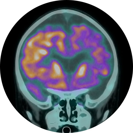
A FDG brain PET/CT is used to image the metabolic function of the brain. Positron emission tomography (PET) uses small amounts of radioactive materials called radiotracers, a special camera, and a computer to help evaluate the brain. It is most commonly used to calculate the degree and pattern of brain volume loss, which allows the neuroradiologist to assess for different types of neurodementia syndromes. It can also help differentiate recurrent brain tumor vs. radiation change after a patient has had brain surgery. Additionally, it can help identify the seizure focus in patients with epilepsy.
icobrain is a very useful quantitative volumetric AI tool that assists neuroradiologists in evaluating patients with dementia, epilepsy, multiple sclerosis, and brain trauma by calculating the volume of different substructures of the brain as well as brain lesions (such as white matter disease or plaques). It compares the volumes to a large normative age- and gender-matched database. This allows the neuroradiologist to know whether the degree of regional brain volume loss is more advanced than expected for the patient's age, which can aid in the diagnosis of a neurodementia syndrome (such as Alzheimer's disease) or other important medical conditions. In multiple sclerosis patients, icobrain calculates the volume and location of intracranial plaques. The software highlights new, enlarging, or shrinking plaques. It is helpful for tracking the rate of disease progression over time. icobrain has both CT and MRI applications. icobrain products in development include volumetric tumor assessment, brain myelination, assessment of brain metabolites, brain perfusion, and diffusion. icompanion is a free health app to support people with multiple sclerosis including symptom tracking, treatment reminders, and image review.
NeuroQuant (NQ) is an artificial intelligence (AI) tool that calculates the volume of different substructures of the brain and compares those to a large normative age- and gender-matched database to determine whether the degree of brain volume loss is statistically significant for patient age. This can be used to improve the early detection of Alzheimer's Disease (AD) or other neurodementia syndromes. NeuroQuant-MS is used to calculate the volume, number, and location of plaques in patients with multiple sclerosis. The software highlights new, enlarging, or shrinking plaques. This allows for accurate tracking of disease status over time. NQ can also be used to detect the location of a seizure focus in patients with epilepsy. It is also used in brain trauma patients or to assess brain development. Neuro-Quant has both MRI and CT applications. Other NeuroQuant-based AI tools in development include volumetric quantification and characterization of brain tumors.
SubtleMR is an AI tool that uses deep learning to allow an MRI of brain, spine (or any other body region) to be acquired 40-60% faster, but with higher image quality than standard scans. This is an exciting new tool that is revolutionizing MR imaging. Having faster and superior quality imaging is appealing to patients, neuroradiologists, and imaging enterprises in general.