

We have a “deep bench” of expertise in musculoskeletal imaging, which is integral for pain management, diagnosis, and treatment of sports-related injuries. Individuals benefit from the same level of professionalism and quality we extend to many professional, amateur, and collegiate teams.
Some Sports Imaging Procedures Include:
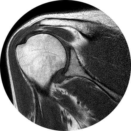
An arthrogram is an X-ray exam of a joint, using a contrast agent and fluoroscopy (a live motion X-Ray). It is used to diagnose the cause of pain or restricted motion of a joint as well as injury to the components of the joint including, the tendons, soft tissues, ligaments, labrum, cartilage and bones. Often this procedure is used to image the shoulder and hip joints, and it is also used when investigating the knees, elbows, ankles and wrists. CT and MRI may also be used to gain additional images of the joint.
X-ray is the oldest and most economical form of medical imaging. During the procedure, radiation passes through the body onto “film” (now digitized and displayed on a computer screen). In neuroimaging, spinal X-rays are used to assess for the degree of spinal motion with flexion or extension.
Myelography is an exam in which contrast material is injected into your spinal column and then that contrast and spinal anatomy is imaged with CT technique. This allows the neuroradiologists to evaluate areas of nerve root impingement, canal narrowing, or disc protrusions. It is typically used when a patient is not a candidate for MRI.
A three phase bone scan is a nuclear medicine test, it uses radiotracers that are injected (or inhaled or swallowed). The radiotracers are detected by a special camera to provide pictures to diagnose a fracture when it cannot be seen on an X-Ray. It is also used to diagnose bone infection, bone pain, osteomyelitis, as well as other bone diseases.