

Description for Alzheimer’s Evaluation.
Description for Amyloid Brain Imaging.
Amyloid brain PET/CT is used to directly image amyloid deposition in the brain which can be found in patients with Alzheimer’s Disease. This can help confirm or exclude this diagnosis in patients with memory loss. Amyloid PET can be positive in patients with Alzheimer’s up to 20 years before the patient becomes symptomatic.
A FDG brain PET/CT is used to image the metabolic function of the brain. Positron emission tomography (PET) uses small amounts of radioactive materials called radiotracers, a special camera, and a computer to help evaluate the brain. It is most commonly used to calculate the degree and pattern of brain volume loss, which allows the neuroradiologist to assess for different types of neurodementia syndromes. It can also help differentiate recurrent brain tumor vs. radiation change after a patient has had brain surgery. Additionally, it can help identify the seizure focus in patients with epilepsy.
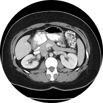
The biliary system is comprised of the liver, gallbladder and bile ducts, and a hepatobiliary exam, or HIDA, evaluates issues in that system. A radioactive tracer is injected (or inhaled or swallowed) and then taken up by the target organs. In this way, the clinician can see how well they function. It shows if the bile ducts are closed or leaking, or if the gallbladder is inflamed, or if the liver is abnormal. HIDA is a nuclear medicine test, which delivers information that often cannot be obtained by other procedures.
A ventilation/perfusion lung scan (also known as a V/Q lung scan) assesses the circulation of air (ventilation) and blood (perfusion) in a patient’s lungs. The exam is useful for identifying blood clots or abnormal flow in the lungs or serious lung disorders such as chronic obstructive pulmonary disease (COPD) or a pulmonary embolism. The V/Q scan uses intravenous material (radiotracers) to show the functional health of the organs.
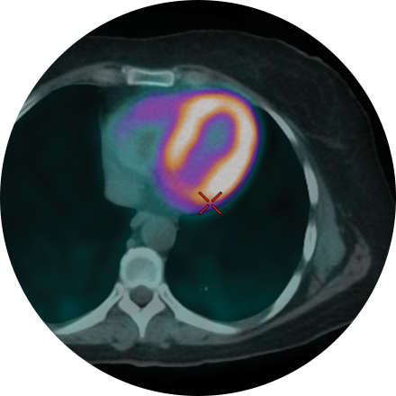
A multi-gated acquisition (MUGA) scan creates video images that show whether the lower chambers (ventricles) of the heart are pumping blood properly. MUGA uses intravenous material (radiotracers) to show how blood moves through the heart. MUGA can be used to check for pre-existing cardiac conditions prior to chemotherapy, or after treatment to assess possible side effects.
Positron emission tomography (PET) and computed tomography (CT) are state-of-the-art diagnostic imaging tools. A PET/CT scan with a sodium fluoride injection is an imaging test that scans the entire skeletal system and produces images of the bones. These images are used to detect areas of abnormal bone growth associated with tumors that may have spread from different parts of the body.
Description for Soft Tissue Imaging.
A three phase bone scan is a nuclear medicine test that uses radiotracers, which are injected. The radiotracers are detected by a special camera to provide pictures to diagnose a fracture when it cannot be seen on an X-Ray. It is also used to diagnose bone infection, bone pain, osteomyelitis, and other bone diseases.
A thyroid scan shows, in video images, how well a patient’s thyroid is functioning, along with its structure and position. This type of study can be used to diagnose hyperthyroidism, cancer, and other abnormalities, such as lumps or inflammation, in this important organ. Like other scans that are used to determine whether an organ is functioning properly, a thyroid scan is a nuclear medicine test; that is, it uses intravenous radiotracers detected by a special camera to provide pictures that show
This exam is most commonly used for diagnosing metastatic disease or primary bone cancers. It may also be used to diagnose Paget’s disease, fractures (old vs. new or post trauma), unexplained bone pain, and other bony abnormalitites.
The procedure itself is non-invasive and simple. The patient receives an intravenous injection of a radiotracer. There are virtually no side effects or allergic reactions to this or any other radiopharmaceutical. The patient may leave after injection and return approximately 3 hours later for the scan. You’ll be asked to lie still on a table while an armlike device supporting a tracer-sensitive camera passes back and forth over your body. Patients are encouraged to stay well hydrated to improve the images. Otherwise they may resume their normal routine. The whole body bone scan takes approximately 45 minutes to one hour for most patients.
Because the radiation dose comes from the injection not the camera, additional images may be obtained of any area of interest, sites of pain, etc. without additional exposure for the patient.