

Nuclear Medicine Services in Annandale, VA
Woodburn Nuclear Medicine & Metro Region PET Center offers a full range of outpatient nuclear medicine studies for patients throughout Annandale, Fairfax, Falls Church, Arlington, and the greater Northern Virginia and Washington, DC metro area. Nuclear medicine scans record images of organs and tissues following the absorption of a small, safe amount of a radiotracer in the body. The images are obtained by a special camera that detects radioactivity, making nuclear medicine unique in its ability to document both organ function and structure. It is used in the diagnosis, management, treatment, and prevention of serious disease.
Below are the nuclear medicine studies we perform most frequently. Looking for PET/CT imaging specifically? Visit our PET/CT Services page.
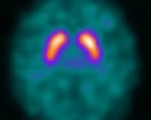
DaTscans
A DaTscan utilizes a radiopharmaceutical to assist in the evaluation of adult patients with suspected parkinsonian syndrome.
Preparation: Some medications or supplements may interfere with this test. Please inform our office of which drugs or supplements you are currently taking.
The total time commitment for this study is approximately 5 hours.
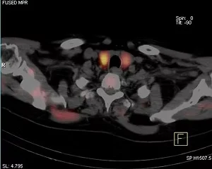
Parathyroid Scan
A parathyroid scan is performed to detect and localize parathyroid adenomas.
Preparation: There is no preparation required for this study.
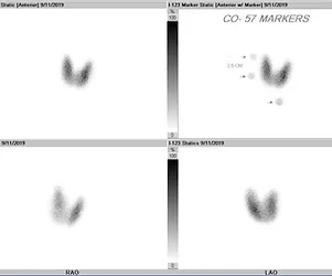
Thyroid Scan, Uptake & Ablation Therapy
A thyroid scan provides information about the thyroid gland and aids in evaluating whether the gland is hyper or hypo active. It is also utilized to assess thyroid cancer.
Thyroid cancer patients should begin a low iodine diet 2 weeks prior to the study and lab results (TSH and serum thyroglobulin) should be made available. On the day of the thyroid scan, patients should have nothing by mouth for 4 hours prior to their appointment time.
I-123 Whole Body Scan with Thyrogen — The patient will receive 2 intramuscular injections of Thyrogen. The injections will be administered on two consecutive days, usually Monday and Tuesday. An I-123 capsule will be administered orally on Tuesday afternoon and the patient will return on Wednesday for imaging. If therapy with I-131 (ablation) has been prescribed, the I-131 capsule will be administered orally on Wednesday after the scan. Post ablation imaging will be scheduled for 8-12 days later and requires no additional preparation.
I-131 Therapy with Thyrogen — Iodine-131 (I-131) is a radioactive form of iodine that is used to destroy (ablate) malignant or hyperactive thyroid tissue. This process requires a time commitment of approximately 60 minutes. The patient will receive 2 intramuscular injections of Thyrogen. The injections will be administered on two consecutive days, usually Monday and Tuesday. An I-131 capsule will be administered orally on Wednesday. Post therapy imaging will be scheduled for 8-12 days later and requires no additional preparation.
Note: If a scan or ablation therapy is being performed after Synthroid withdrawal, Thyrogen will not be administered. The whole body scan will be performed 24 hours after I-123 administration.
Thyroid Scan & Uptake with I-123 — Patients should avoid seafood for 3 days prior to the scan and the patient should not receive iodinated contrast for 6 weeks prior to the test. Also, the patient should not be on thyroid replacement (Synthroid) or anti-thyroid medication (e.g. Tapazole or PTU) at the time of the test.
Download the I-131 Therapy Brochure Download the Thyroid Scan Brochure
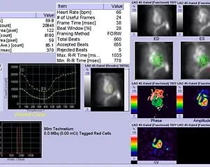
MUGA Scans (Rest)
MUGA stands for multi-gated acquisition. This nuclear medicine test is utilized to measure the ability of the heart to contract. A tiny amount of blood is removed and labeled with a radioactive material called Technetium. The labeled red blood cells are then injected back into the patient and a scanner takes pictures of the heart contracting. Also, the electrical activity of the heart is measured during this scan.
Preparation: No preparation is required for this test.
Please allow 1-2 hours for this test.
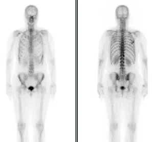
Bone Scans
Bone scans are a sensitive test for evaluating almost any process affecting the skeleton, like bone tumors, fractures, infection, inflammation and many other conditions. The study may be abnormal days or weeks before there are symptoms or changes on plain x-rays. The study is usually completed at one visit, requiring anywhere from one to several hours depending on the reason for the study. SPECT imaging can be performed to give three-dimensional imaging that can be compared directly to images from a CT or MRI. Serial examinations over months or years can document the extent or severity of a disease process over time.
Preparation: For all types of bone scans, the patient should drink as much fluid as possible before and after the procedure. A total of 32 ounces is recommended.
The total time commitment for this study is approximately 4 hours.
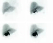
Hepatobiliary (HIDA) Scan
A HIDA scan is a nuclear medicine test which examines the flow of bile as it is produced in the liver and travels into the gallbladder and small intestine. Gall bladder inflammation (cholecystitis) can prevent the gallbladder from filling with bile or emptying correctly. A HIDA scan can test for abnormal gallbladder function as a cause of abdominal pain.
Preparation: Please have nothing to eat or drink for at least 4 hours prior to your appointment time.
Please allow for 1 1/2 - 2 hours for this scan.
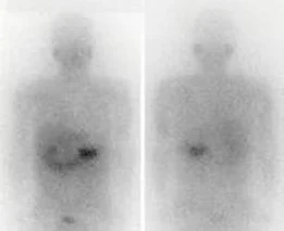
I-123 MIBG
This study requires injection of the radiopharmaceutical I-123 MIBG. The radiopharmaceutical will identify neuroendocrine tumors with the use of a gamma camera. Imaging is performed at 24 hours post injection and occasionally at 48 hours. A number of medications may interfere with this test so the patient may be asked to delay or discontinue some of them prior to the test. A complete medication list should be provided at the time of the scan.
Labeled White Blood Cell (WBC) Scan
A Labeled WBC Scan is a nuclear medicine procedure in which white blood cells are removed from the patient, tagged with the radioisotope Indium-111 and then injected intravenously into the patient. The tagged white blood cells then localize to areas of relatively new infection.
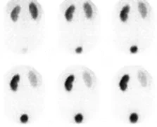
Renal Scan
A renal scan is a nuclear medicine exam in which a small amount of radioactive material is used to measure the function of the kidneys, evaluate renal perfusion, detect obstruction, etc. The specific type of renal scan performed will be determined by the patient's indication and referring physician's order.
Preparation: Patients should be well-hydrated by drinking at least 32 ounces of fluid prior to their appointment. Additional preparation will vary depending on the specific type of renal scan to be performed. Our staff will discuss the patient's preparation requirements during scheduling.
The total time commitment for this study is approximately 2 hours.
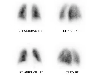
Pulmonary Lung Scan (V/Q and Quantitative)
Ventilation and perfusion lung scans are performed to analyze the flow of blood and movement of air into and out of the lungs. It is used most often to detect pulmonary emboli or blood clots in the lungs.
Preparation: No preparation is required for these tests.
The total time commitment for this study is approximately 1 hour.
Why Choose Woodburn for Nuclear Medicine in Northern Virginia
- Full range of outpatient nuclear medicine studies in one Annandale location
- Experienced nuclear medicine technologists focused on patient comfort and safety
- Convenient access near Fairfax, Vienna, Merrifield, Centreville, Manassas, Springfield, Woodbridge, Alexandria, and Washington, DC
- Collaboration with referring physicians for timely, detailed imaging reports
- ACR Accredited facility
Need PET/CT imaging for oncology or neurological evaluation? Visit our PET/CT Services page.
Schedule a Nuclear Medicine Study
Nuclear Medicine Phone: (703) 698-0666
3289 Woodburn Road, Suite 060
Annandale, VA 22003