
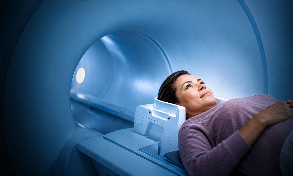
What is an MRI?
MRI is a special diagnostic test that produces very clear, detailed pictures of internal organs and structures in your body.
It uses a magnetic field, radio waves and a computer to create cross-sectional images — slices through the body that can be viewed from any angle. Because it reads soft tissue so well, MRI can show some problems that other exams may miss.
When is it used?
Your physician may order an MRI to look closely at soft tissue, the nervous system, or an injury that other imaging cannot fully explain.
-
01
Brain and spine
Identifying stroke, multiple sclerosis and spinal disc herniations, with quantitative brain volumetry to help evaluate neurodegenerative conditions.
-
02
Tumors
Visualizing tumors and defining their size, position and relationship to nearby tissue.
-
03
Joints
Evaluating joint problems, and injuries to the knee, shoulder, back or neck.
-
04
Soft tissue
Diagnosing soft tissue conditions that show poorly on X-ray or CT.
-
05
Eyes and ears
Identifying diseases affecting the eye and the inner ear.
-
06
Injury
Assessing damage after an injury, when the extent is not clear from other imaging.
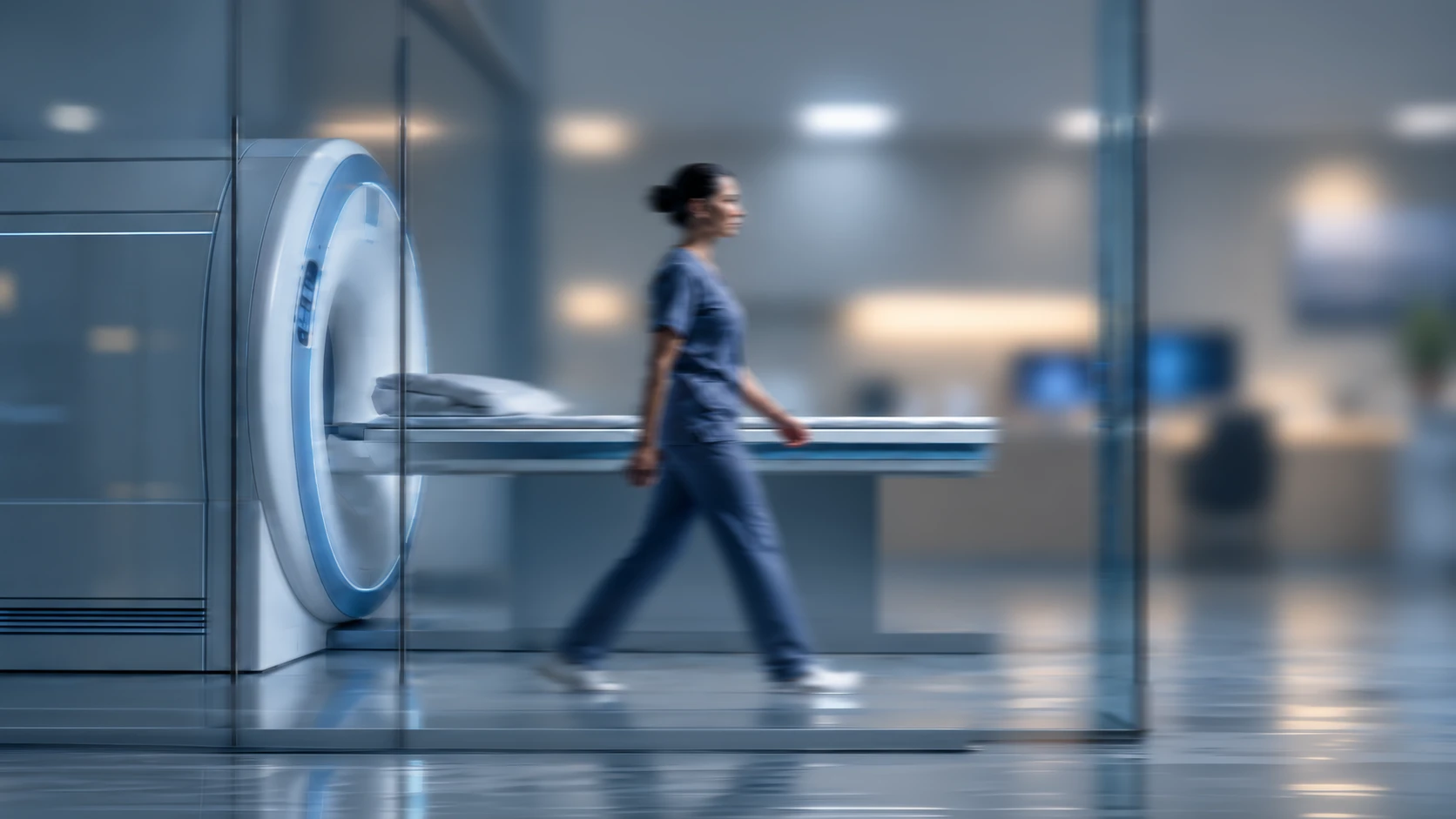
Inside the exam
You lie down
You rest on a cushioned bed that moves slowly into the scanner. Your technologist can see you and talk with you the whole time.
Contrast, if needed
Some exams use a contrast medium called gadolinium, injected into a vein, to make certain tissues clearer.
It gets loud
Knocking, buzzing, a rhythm that changes as the scan moves through its sequences. That sound is the machine building your images. You will have earplugs or headphones.
A radiologist reads it
A contracted radiologist reviews your images and sends a report to your physician. Urgent findings are communicated right away.
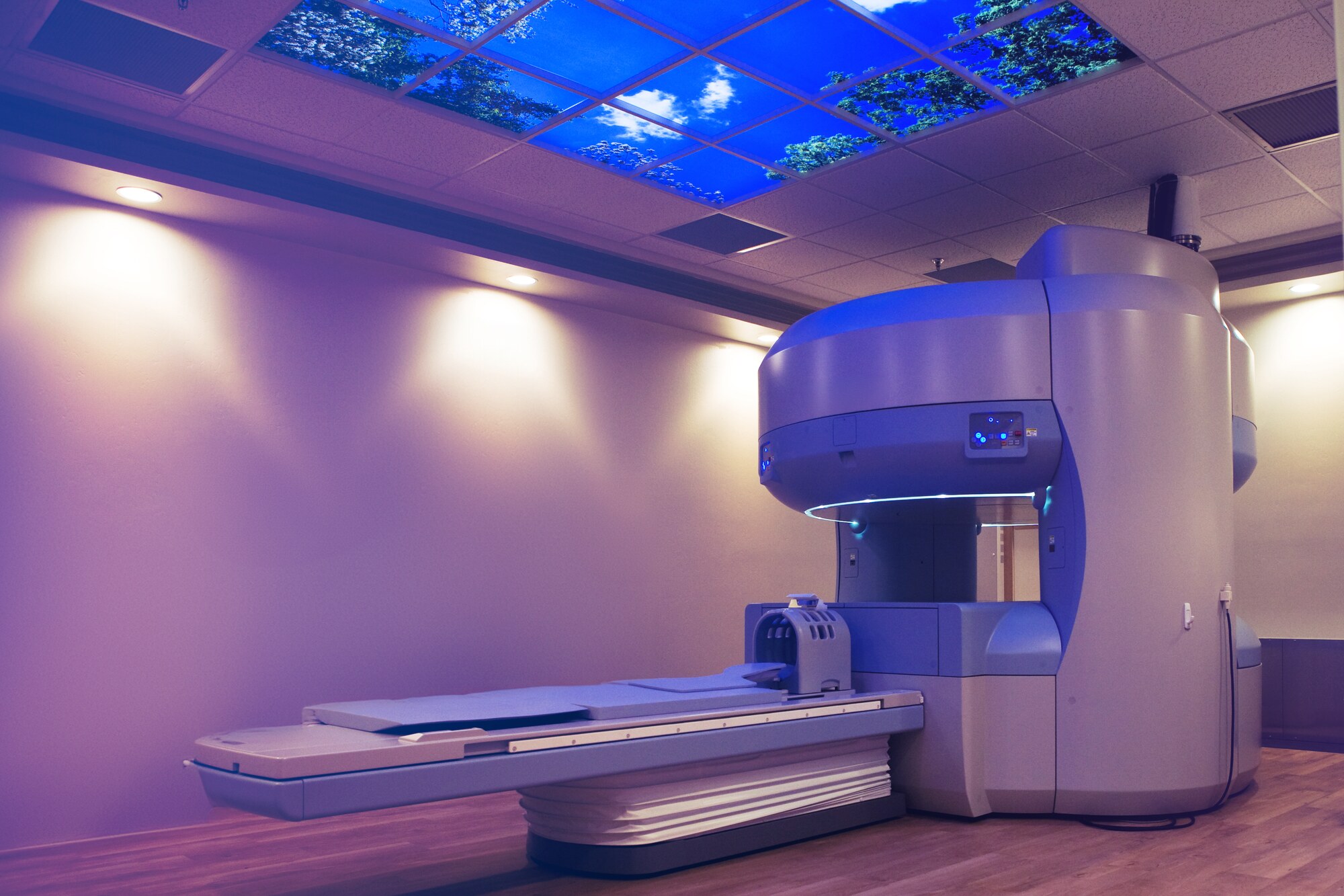 Pictured: Kern Radiology · Bakersfield, CA
Comfort during your MRI
Around 30% of people have an anxious or claustrophobic reaction to a traditional MRI. A lot of them put the exam off because of it, and never mention it. Tell us when you book. It changes which scanner we put you on, not whether you can have the exam.
Wide-Bore, Wide-Open and Open MRI
RadNet offers Wide-Bore, Wide-Open, and Open MRI configurations alongside AI-accelerated faster sequences, giving you extra space and significantly shorter scan times. The Wide-Bore scanners take patients up to 500 pounds.
Exam preparation
We want your exam to be as comfortable and hassle-free as possible, so we provide all the information you will need to prepare before you arrive.
Your specific exam has its own instructions. You will find them on the Exam Preparation page.
All exam preparationWhat to bring
-
Prescription or referral from your physician.
-
A list of the medications you take, including non-prescription medications and supplements.
-
Insurance cards.
-
Any previous, relevant imaging exams and reports performed outside of our network, and your medical history.
Common questions
Yes. MRI uses magnetic fields and radio waves rather than ionizing radiation, so there is no radiation exposure. Most MRI exams are painless, and the exam poses almost no risk to the average patient when appropriate safety guidelines are followed.
Most MRI exams take 30 to 60 minutes. RadNet integrates deep-learning AI reconstruction to accelerate scan times by 40–60%, maximizing image clarity while minimizing time spent inside the scanner.
The knocking and buzzing is the sound of the gradient coils switching on and off as they build each image. It is normal, it means the scanner is working, and you will be given earplugs or headphones.
It depends on what the metal is and where it sits. Many implants can be scanned safely under specific conditions, and some cannot. Tell us about any implant, stent, surgical clip, pacemaker or metal fragment when you schedule, so we can check it before your appointment rather than on the day.
RadNet offers Wide-Open and Open MRI scanners at many locations. Tell us when you schedule and we will book you at a center with the scanner that suits you best.
Some exams use a contrast medium called gadolinium, injected into a vein, to make certain tissues clearer. Your physician decides whether contrast is needed when the exam is ordered.

Find an MRI location near you
Scanner types and available exams vary by center. Confirm the exam you need when you book.