
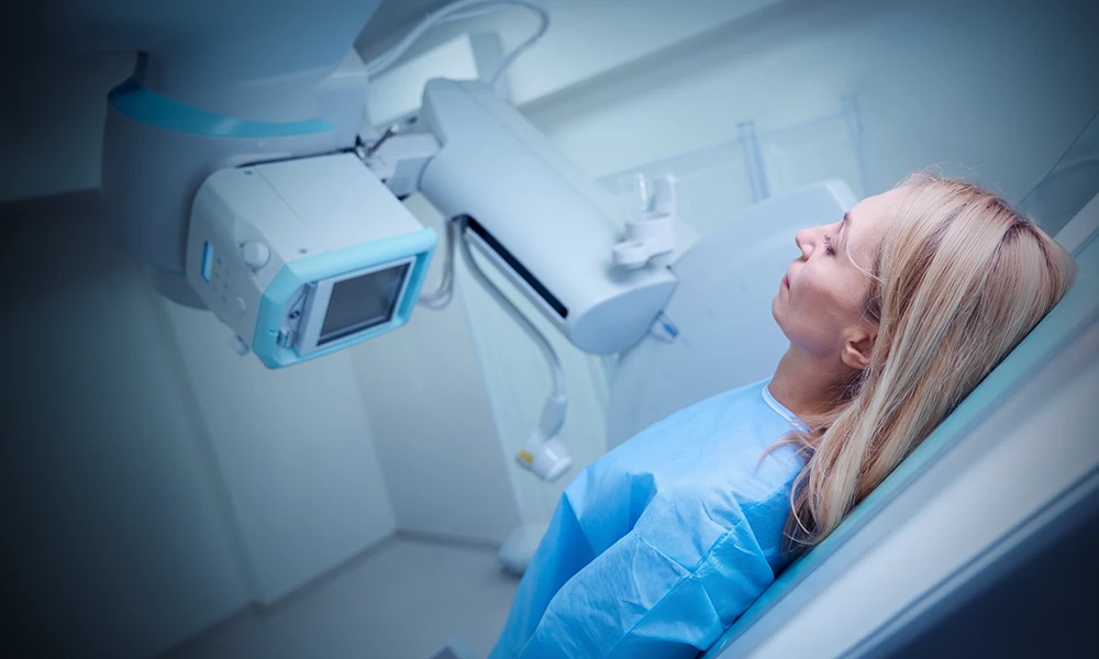
What is fluoroscopy?
Fluoroscopy uses a continuous X-ray beam to produce images of organs and bones in real time, on a television monitor or screen, like an X-ray movie.
Contrast material
During this procedure, radiologists usually use contrast material (dye) to highlight the area being examined.
The contrast material can be injected or taken orally or rectally, depending on the area being examined.
How fluoroscopy differs from an X-ray
An X-ray takes one picture. Fluoroscopy keeps the X-ray beam on, so your physician can watch the movement happen instead of reading it afterwards.
A barium swallow. Because the beam stays on, the contrast is seen traveling — a still X-ray would catch it at one point on the way down, and nothing either side of that moment.
When is fluoroscopy used?
Some studies use fluoroscopy to watch an organ work. Others use it to guide a clinician’s hand while they work.
-
01
The gastrointestinal tract
Investigations of the gastrointestinal tract. A barium enema for the colon is one example.
-
02
Barium swallow and upper GI
Evaluating swallowing mechanics, esophagus, stomach, and reflux.
-
03
Joint injections and arthrograms
Guiding precise needle placement for therapeutic pain injections or joint MRI prep.
-
04
Hysterosalpingography (HSG)
Evaluating uterine structure and fallopian tube patency for fertility assessments.
-
05
Myelography
Visualizing the spinal cord and nerve roots when MRI is contraindicated.
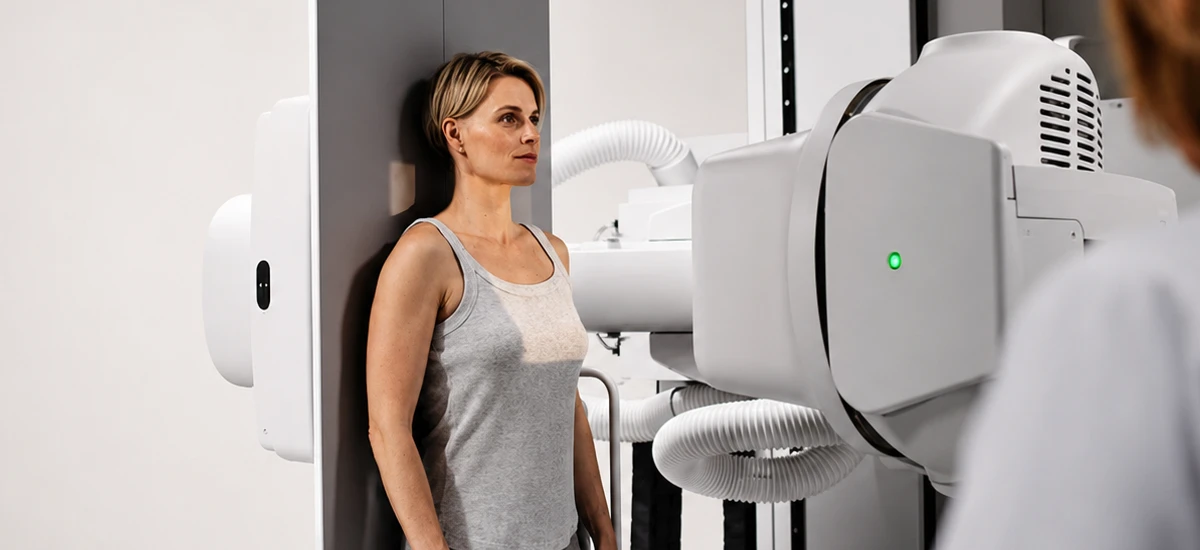
Inside the exam
The X-ray itself is painless. Some fluoroscopy exams use a needle, catheter or tube, which can cause brief discomfort.
Fluoroscopy helps diagnose diseases and guide clinicians during treatment, and we use the lowest acceptable exposure for the shortest time necessary in order to reduce your risk.
During the exam
-
The technologist will position you sitting, standing, or lying on the exam table, and then conduct the procedure.
-
You will be able to communicate with the technologist during the procedure.
Afterwards
-
Most patients can leave right after the exam. Your care team will tell you if you need to stay for observation or need someone to drive you home.
-
Unless your doctor told you to limit fluids, drink plenty of fluids, especially water, for the next 24 hours to help flush the contrast material from your body.
About the radiation
Digital images and flat-panel detector systems reduce your exposure to radiation during this procedure, and the amount of exposure depends on the particular type of fluoroscopy.
Exam preparation
We want your exam to be as comfortable and hassle-free as possible, so we provide all the information you will need to prepare before you arrive.
Your specific exam has its own instructions. You will find them on the Exam Preparation page.
All exam preparationWhat to bring
-
Prescription or referral from your physician.
-
A list of the medications you take, including non-prescription medications and supplements.
-
Insurance cards.
-
Any previous, relevant imaging exams and reports performed outside of our network, and your medical history.
Common questions
Fluoroscopy uses a continuous X-ray beam to produce images of organs and bones in real time, on a television monitor or screen, like an X-ray movie.
The X-ray itself is painless. Some fluoroscopy exams use a needle, catheter or tube, which can cause brief discomfort. The technologist will position you sitting, standing, or lying on the exam table, and you will be able to communicate with the technologist during the procedure.
During this procedure, radiologists usually use contrast material (dye) to highlight the area being examined. The contrast material can be injected or taken orally or rectally, depending on the area being examined.
Digital images and flat-panel detector systems reduce your exposure to radiation during this procedure, and the amount of exposure depends on the particular type of fluoroscopy. We use the lowest acceptable exposure for the shortest time necessary in order to reduce your risk.
Most patients can leave right after the exam. Your care team will tell you if you need to stay for observation or need someone to drive you home. Unless your doctor told you to limit fluids, drink plenty of fluids, especially water, for the next 24 hours to help flush the contrast material from your body.
Specific fasting instructions apply for Upper GI or Barium studies. Please confirm these requirements with our scheduling team to ensure your study can be completed as planned. Different radiology exams have different preparation requirements, and our staff will discuss specific instructions with you.
If you are pregnant, or think you might be pregnant, let your doctor know. Your doctor may go ahead with, change or postpone the exam. A needed X-ray can still be done.

Find fluoroscopy near you
Fluoroscopy is not offered at every center. Confirm the exam you need when you book.