
What is an X-ray?
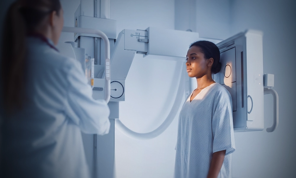
X-rays can easily pass through body tissue, making it possible to provide images of internal structures without performing surgery.
During the procedure, electromagnetic radiation passes through the body onto film — now digitized and displayed on a computer screen. Dense structures such as bone absorb most of the radiation and appear white on the digital image. Structures that are less dense, like air, appear black. Everything in between appears a different shade of gray.
X-ray can also help evaluate and monitor some diseases, including certain heart, lung and bone conditions and some cancers. Osteoporosis is diagnosed with a bone density (DEXA) scan.
When is it used?
Digital X-rays are used to diagnose a wide range of illnesses and injuries.
It is the fastest, easiest and most economical way for a physician to view and assess broken bones, and it is used to diagnose and monitor the progression of disease over time.
-
01
Musculoskeletal injuries
-
02
Cancer
-
03
Chest and cardiopulmonary conditions
Evaluating pneumonia, congestive heart failure, fluid accumulation, and chronic lung disease.
-
04
Abdominal pain
-
05
Sinus disease
-
06
Spinal problems
How do I get an X-ray?
X-ray remains the foundation of medical imaging, providing rapid answers for bone health, chest conditions and emergency diagnostics. With the largest network of outpatient imaging centers in the country, RadNet provides walk-in access for standard X-ray services.
A second set of eyes
At participating centers, X-ray reads use DeepHealth’s AI fracture detection, which flags possible fractures for the radiologist to review before the final report.
Why digital?
Digital imaging gives us many advantages in handling your exam, not the least of which is faster communication of results to your doctor. It also allows your medical team to collaborate, and to immediately compare previous exams with current ones, so that your health is properly monitored.
CT and MRI
Contracted radiologists use X-rays to provide a clear image of the bone, and to see where and what type of fracture is present. But X-rays do not show soft tissues like ligaments, tendons and cartilage well. If the injury is more severe, contracted radiologists may use an MRI or a CT scan.
CT
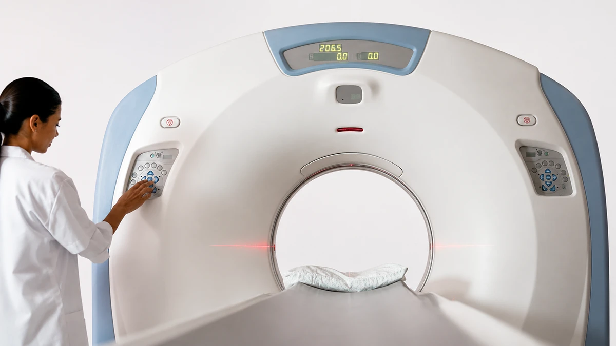
Cross-sectional images that show a complex or severe fracture in detail an X-ray cannot resolve.
Explore CTMRI
The soft tissue an X-ray shows poorly: ligaments, tendons and cartilage around the injury.
Explore MRIExam preparation
We want your exam to be as comfortable and hassle-free as possible, so we provide all the information you will need to prepare before you arrive.
For an X-ray there is genuinely very little to do. Bring what is on the list, and tell us if there is any possibility you are pregnant.
All exam preparationWhat to bring
-
Prescription or referral from your physician.
-
A list of the medications you take, including non-prescription medications and supplements.
-
Insurance cards.
-
Any previous, relevant imaging exams and reports performed outside of our network, and your medical history.
Common questions
No. The X-ray itself is painless, though holding a position or the cool room can be uncomfortable. It usually needs no preparation, and a radiologist reads the images and sends a report to your doctor.
This exam requires no special preparation. Bring your prescription or referral, a list of your medications, your insurance cards and any previous relevant imaging.
Many centers take walk-in X-rays with a physician’s order; hours vary by center. RadNet has the largest network of outpatient imaging centers in the country, so there is usually a center close to you.
A digital X-ray uses a small amount of radiation, and the cancer risk from it is generally very small.
Always inform the technologist if there is any possibility of pregnancy prior to the exam. If you are pregnant, or think you might be pregnant, let your doctor know. Your doctor may go ahead with, change or postpone the exam. A needed X-ray can still be done.
You may be asked to stand or lie down on an examination table, depending on the part of the body to be examined. You will be able to communicate with the technologist during the procedure.
Cost depends on the exam and on your insurance. Ask our scheduling team about cost when you schedule, and you can request a Good Faith Estimate if you are paying without insurance.

Find an X-ray location near you
Not every center offers every X-ray exam. Confirm the exam you need when you book.