
About the fellowship
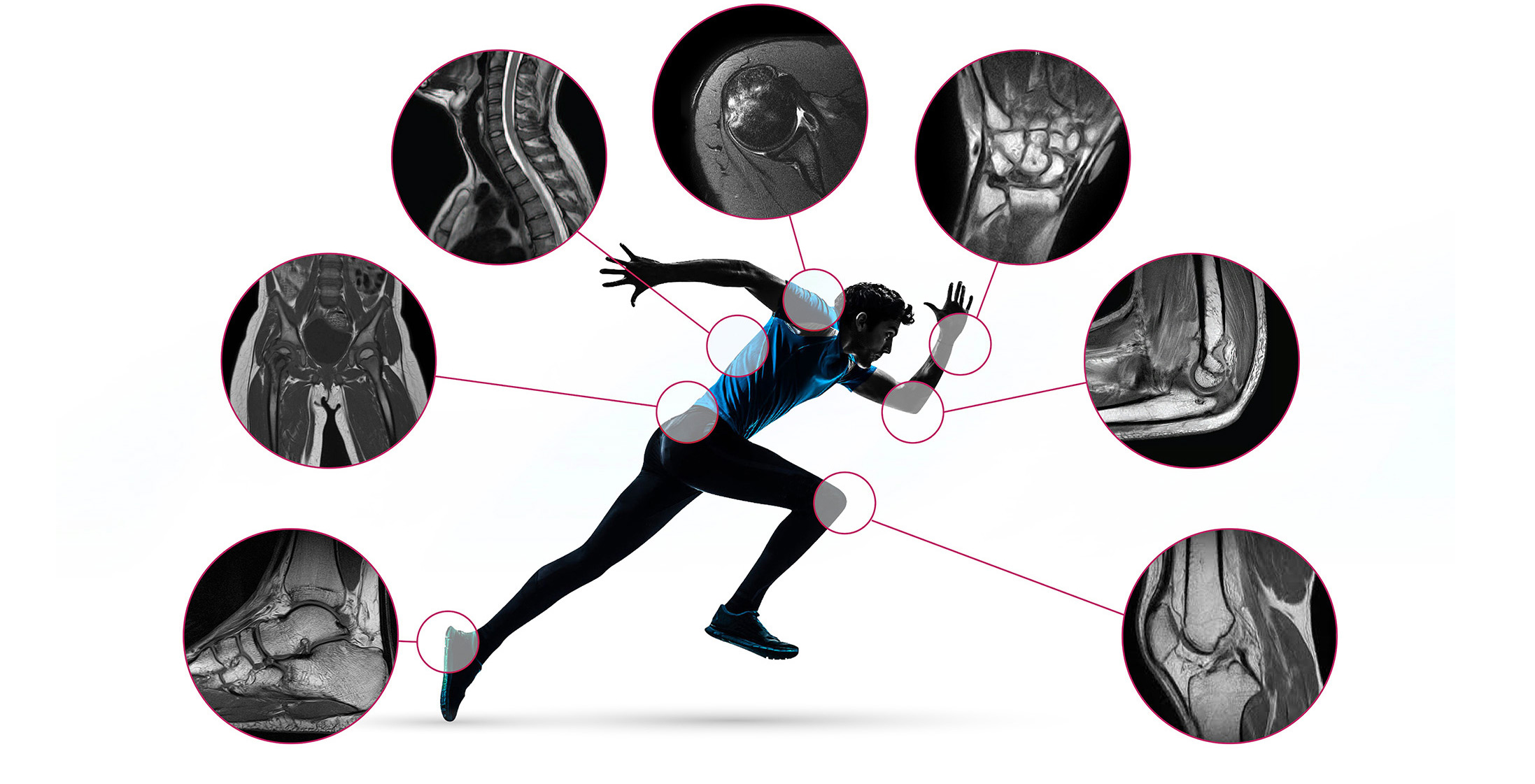
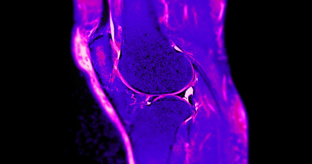
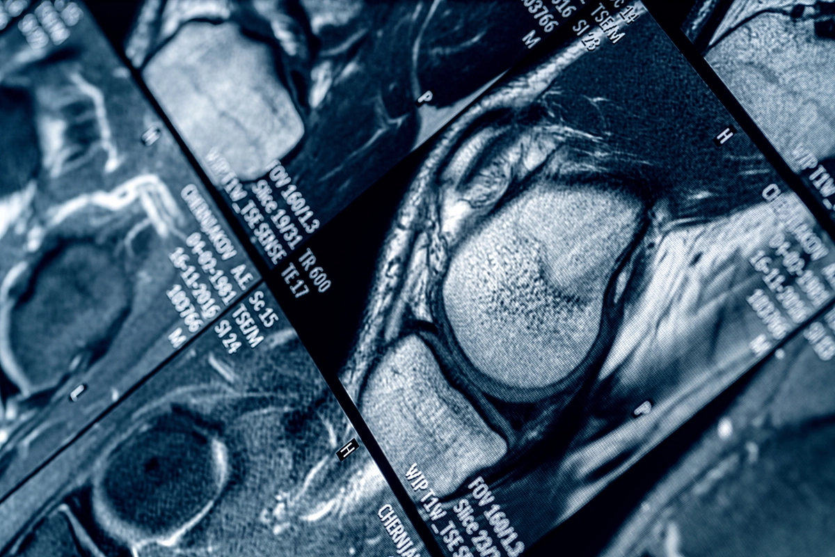
Fellows gain extensive experience in advanced musculoskeletal imaging, with a high volume of MRI and CT examinations spanning the full spectrum of orthopedic and sports-related pathology.
Our close relationships with premier orthopedic groups and nationally recognized sports medicine programs give fellows a unique perspective on imaging as an integral part of patient care — from diagnosis and treatment planning through postoperative evaluation and return-to-play decisions.
The program’s affiliations with professional, collegiate and Olympic sports offer a valuable opportunity to meet the demands of elite athletic medicine and the advanced imaging required to care for high-performance athletes.
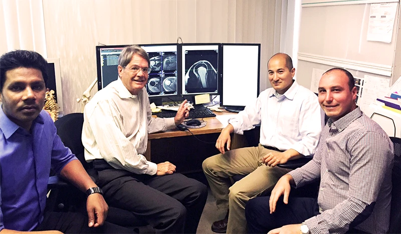
Fellows work closely with recognized leaders and accomplished radiologists in musculoskeletal imaging, gaining individualized mentorship and exposure to diverse approaches to diagnosis, interpretation, and clinical decision-making. Regular didactic MSK lectures, participation in sports medicine and orthopedic lectures, and side-by-side readouts are all part of the curriculum, along with instruction in outpatient image-guided MSK procedures. The emphasis is not simply on reading studies, but on developing the expertise, confidence, and judgment required to practice at the highest level of MSK radiology.
Excellence in medicine should coexist with a fulfilling life outside the reading room. Our Southern California setting offers an extraordinary lifestyle, while the fellowship emphasizes work-life balance, collegiality, and a supportive learning environment.
Fellows here are not simply preparing for their next job. They are developing the expertise, professional relationships, and clinical perspective to become the next generation of leaders in musculoskeletal radiology.
Program highlights
- High-volume advanced imagingExtensive exposure to sophisticated MRI and CT cases covering sports, orthopedic, rheumatologic, trauma, and oncologic pathology.
- Elite sports medicine affiliationsExposure to, and imaging experience with, professional, collegiate and Olympic athletes.
- Integrated clinical careClose partnerships with premier orthopedic and sports medicine groups, focused on treatment planning, postoperative evaluation, and return-to-play decisions.
- Dedicated mentorshipDaily one-on-one readout and case review alongside nationally recognized MSK radiology leaders.
- Southern California lifestyleA supportive, collegial environment in one of the most vibrant regions in the country.
The program
RadNet and the fellowship
A high volume of musculoskeletal MRI cases are drawn from multiple sites in the RadNet and Pronet teleradiology networks. This includes multiple orthopedic/sports medicine and rheumatology groups. The fellow will gain experience with high field, 1.5/3.0 Tesla MR studies as well as low-field 0.2 to 1.0 T extremity scanners (E-Scan and C-Scan, ONI, MagneVue).
Diverse cases
Additional experience with Neuroradiology, Abdominal Imaging, Breast MRI, and PET/CT Imaging depending on degree of personal interest.
Academics
Dedicated academic time is encouraged for performing clinical research. Fellows are encouraged to submit at least one paper to a peer-reviewed journal during their year of training.
Lectures
Daily lectures given by Dr. Crues covering MSK-MRI and MRI physics throughout the year.
Conferences
Weekly multidisciplinary conferences at Cedars-Sinai Orthopaedics.
Cedars-Sinai Orthopaedics
Affiliation with Cedars-Sinai Orthopaedics and the Cedars-Sinai Orthopaedic Sports Medicine Fellowship, including shared conferences, and hosting its orthopedic and sports medicine fellows for educational sessions and MSK radiology readouts.
Requirements
The regular one-year MSK fellowship requires that fellow applicants have a valid U.S. Medical License at the time of application and a valid California Medical License before starting the program.
Visiting fellowships
Visiting fellowships are available for between 1-6 months for non-U.S. radiologists by special arrangement with Dr. Lee.
Outside rotations
Joint arthrography
Diagnostic and therapeutic injection of ankle, knee, hip, spine, wrist, elbow, and shoulder.
The Stone Clinic · San Francisco
Clinical evaluation of orthopedic and sports medicine conditions. Observation of orthopedic and arthroscopic procedures, and conducting collaborative research projects.
Elective rotations
Elective rotations in breast, neuro, body and PET imaging are available.
Affiliations
- Cedars-Sinai Orthopaedics and the Cedars-Sinai Orthopaedic Sports Medicine Fellowship
- Southern California Orthopedic Institute
- The Stone Clinic
- Santa Barbara Cottage Hospital Radiology Residency Program
- Riverside Community Hospital Radiology Residency Program
- Society of Skeletal Radiology
- RadNet and Pronet teleradiology networks
- Beverly Radiology Medical Group
Lectures and protocols
Through our Musculoskeletal MRI Fellowship program, participants review both high- and low-field MRI cases together under the direction of Dr. John Crues.
Protocol documents
Click the protocols below to open or download a PDF file.
- MSK Protocols PDF
- Neuro Protocols PDF
- Body MRI Protocols PDF
- Body MRI Protocols PDF
The above protocols are subject to change and updates.
Fellowship publications
RadNet musculoskeletal fellows have been actively involved in publishing scientific and educational articles. Some of these are listed below.
-
01
Variation in glenohumeral rotation affects the ability to identify subcoracoid impingement on magnetic resonance imaging in patients with rotator cuff pathology
The American Orthopaedic Society for Sports Medicine 2012 Annual Meeting, Baltimore, Maryland, July 2012
-
02
Analyzing the Accuracy of Magnetic Resonance Imaging, Arthroscopy, and 3-dimensional Digitalization System in the Evaluation of Meniscal Loss Following Partial Meniscectomy.
The American Orthopaedic Society for Sports Medicine 2012 Specialty Day, San Francisco, February, 2012
-
03
18F PET/CT Imaging of the Spine: Retrospective 3-Month Review of Studies
Presented at the Western Region Society of Nuclear Medicine, Oct. 2010
-
04
Wei, A.S., Khana, S., Limpisvasti, O., Crues, J., Podesta, L., and Yocum, L.A. “Clinical and Magnetic Resonance Imaging Findings Associated with Little League Elbow.” Journal of Pediatric Ortho (2010); 30(7):715-719.
-
05
Yokoo, T., Bae, W.C., Hamilton, G., Karimi, A., Borgstede, J.P., Bowen, B.C., Sirlin, C.B., Chung, C.B., Crues III, J.V., Bradley, W.G., Bydder, G.M. “A Quantitative Approach to Sequence and Image Weighting.” J Comput Assist Tomogr. 2010 May-Jun; 34(3): 317-31. Review.
-
06
Stone, KR., Crues III, J.V., Dang, D., Pelsis JR., Adelson WA., Freyer A., Turek, TJ. “The Use of Articular Cartilage Paste Graft in Massive Articular Cartilage Defects of the Femoral Condyle.” A.N.N.A. Annual Meeting, April 2011 (Submitted).
-
07
Olech, E., Crues, III, J.V., Yocum, D.E., and Merrill, J.T. “Bone Marrow Edema is the Most Specific Finding for Rheumatoid Arthritis (RA) on Noncontrast Magnetic Resonance Imaging of the Hands and Wrists: A Comparison of Patients with RA and Healthy Controls.” J Rheum 2010; 37(2): 265-74.
-
08
Diaz, F.L., Tweardy, L., and Shellock, F.G. “Cervical External Immobilization Devices: Evaluation of Magnetic Resonance Imaging Issues at 3.0 Tesla.” Spine 2010; 35(4): 411-5.
-
09
Eyer B and Crues III JV. “Imaging of the Elbow,” in Sports Medicine of Baseball. Ed. Dines J ElAttrache NS, Yocum LA, Altchek DW, Andrews J, and Wilk KE. Lippincott, Williams & Wilkins, Philadelphia, 2010 (in press).
-
10
Chang J, Crues III JV, Troum OM. “MRI in rheumatoid arthritis: Why start using it now? A review of imaging modalities, MR scoring, and pathophysiology in RA.” Contemporary Topics in Rheumatoid Arthritis 2009; 3(4):2-9.
-
11
Gandhi RT, Kuo R, Crues III JV. “Technical considerations of musculoskeletal imaging at 3.0 tesla.” Seminars in musculoskeletal radiology (2008); 12(3): 185-95.
-
12
Khanna S and Crues III JV. “Complexities of MRI and Other False Positives.” MRI and ultrasound in the diagnosis and management of rheumatological diseases. Annals of the New York Academy of Sciences (accepted).
-
13
Stone KR, Walgenbach AW, Freyer AF, Turek TJ, Crues III JV, Reuter RM. “Salvage and repair of osteochondritis dissecans with articular cartilage paste grafting.” Journal of Bone and Joint Surgery (in press).
-
14
Reuter RM, Shellock FG, Crues III JV, Hiller WD, Ainge G. Brown D., Dierenfield LA. “MRI of the shoulder: Findings in symptomatic and asymptomatic ironman triathletes.” Skeletal Radiology (2008); 37(8): 737-41.
-
15
Frank JB, ElAttrache NS, Dines JS, Blackburn A, Crues JV, Tibone JE. “Repair site integrity after arthroscopic ‘transosseous-equivalent/suture-bridge’ rotator cuff repair.” Am J Sports Med (2008); 36:496-503.
-
16
Stone KR, Freyer A, Turek T, Walgenbach AW, Wadhwa S, Crues J. “Meniscal sizing based on gender, height, and weight.” Arthroscopy. 2007 May;23(5):503-8.
-
17
Furst DE, Gaylis N, Bray V, Olech E, Yocum D, Ritter J, Weisman M, Wallace DJ, Crues J, Khanna D, Eckel G, Yeilding N, Callegari P, Visvanathan S, Rojas J, Hegedus R, George L, Mamun K, Gilmer K, Troum O. “Open-label, pilot protocol of patients with rheumatoid arthritis who switch to infliximab after an incomplete response to etanercept: the opposite study.”
Ann Rheum Dis. 2007 Jul;66(7):893-9. Epub 2007 Apr 5. -
18
Dick, M and Crues JV III. “Bone Deep.” Advance for Imaging and Oncology Administrators. 2007 May 2007 17 (5):41
-
19
Muhammad Ali, MBBS; Tim S. Chen, MD; John V. Crues III, MD. “MRI of the foot.”
Applied Radiology. 35 (12) December 2006 10-20. -
20
Ghazinoor S, Crues III JV, Crowley C. “Low-Field Musculoskeletal MRI.” Journal of Magnetic Resonance Imaging, Special Issue: Frontiers in Musculoskeletal Imaging, February 2007.
-
21
Kuo R, Panchal M, Tanenbaum LN, Crues JV. “3.0 Tesla Imaging of the Musculoskeletal System.” Journal of Magnetic Resonance Imaging, Special Issue: Frontiers in Musculoskeletal Imaging, February 2007.
-
22
Chen TS, Crues JV 3rd, Ali M, Troum OM. “Magnetic resonance imaging is more sensitive than radiographs in detecting change in size of erosions in rheumatoid arthritis.” J Rheumatol. 2006 Oct; 33(10):1957-67. Epub 2006 Aug 1.
-
23
Muhammad Ali, MBBS; Tim S. Chen, MD; John V. Crues III, MD. “MRI of the ankle.”
Applied Radiology. Volume: 35 Number: 8 August 2006 27-39 -
24
Ghazinoor S, Crues JV 3rd. “Low field MRI: a review of the literature and our experience in upper extremity imaging.” Clin Sports Med. 2006 Jul;25(3):591-606, Review.
-
25
Chen TS, Crues JV, Ali M, Troum OM. “Detection of changes in size of erosions in inflammatory arthritis using high resolution in-office MRI.” Proceedings of the International Society for Magnetic Resonance in Medicine, Miami Beach, Fl., May 12, 2005:p133.
-
26
Pouldar D, Matthews R, Rahbar G, Chen E, Jain H, et al. (2016) “Pearls and Pitfalls of 18F Sodium Fluoride PET-CT Bone Imaging for Evaluation of Back Pain and Spine Pathology.” J Nucl Med Radiol Radiat Ther 1: 005.
-
27
Pouldar D, Bakshian S, Matthews R, Rao V, Manzano M, Dardashti S. “Utility of 18F sodium fluoride PET/CT imaging in the evaluation of postoperative pain following surgical spine fusion.” Musculoskeletal Surg. 2017 Aug; 101(2): 159-166.
-
01
MR Arthrogaphy vs. MDCT Arthrogaphy of Hip Labral and Cartilage Pathology
Clinical Magnetic Resonance Imaging, 3rd Ed., Textbook Update. Chapter 97, page 3172, after last paragraph.
-
02
MRI and Radiographic Evaluation of Acromioclavicular Joint Dislocation
Clinical Magnetic Resonance Imaging, 3rd Ed., Textbook Update. Section VII, Chapter 99, page 3267, right after figure 99-85, before “Muscle Injuries” heading..
-
03
Iliopsoas impingement as a potential cause for anterior labral pathology
Clinical Magnetic Resonance Imaging, 3rd Ed., Textbook Update. Chapter 102, page 3398.
-
04
MRI Appearance of Ulnar Collateral Ligament Reconstruction
Clinical Magnetic Resonance Imaging, 3rd Ed., Textbook Update. Section VII, Chapter 100, after page 3307.
-
05
THE POSTOPERATIVE HIP: Imaging of Metal on Metal Prostheses and Their Complications
Clinical Magnetic Resonance Imaging, 3rd Ed., Textbook Update. Section VII, Chapter 102, after page 3399.
-
06
Obesity Prevention and Osteoarthritis Risk Reduction
Clinical Magnetic Resonance Imaging, 3rd Ed., Textbook Update. Chapter 109, page 3587, after last paragraph.
-
07
Trochlear Dysplasia
Clinical Magnetic Resonance Imaging, 3rd Ed., Textbook Update. Section 7, Chapter 103, page 3428, add to last paragraph.
-
08
The Diagnostic Utility of MRI in Spondyloarthritis
Clinical Magnetic Resonance Imaging, 3rd Ed., Textbook Update. Section 7, Chapter 111, page 3868, after 4th paragraph under Rheumatologic Disorders heading.
-
09
Entrapment of the Brachial Plexus
Clinical Magnetic Resonance Imaging, 3rd Ed., Textbook Update. Chapter 75, page 2367 after last paragraph.
-
10
2010 Rheumatoid Arthritis Classification Criteria
Clinical Magnetic Resonance Imaging, 3rd Ed., Textbook Update. Section 7, Chapter 111, page 3636, after 2nd paragraph under Rheumatologic Disorders heading.
-
11
Deep Peronal Nerve Entrapment
Clinical Magnetic Resonance Imaging, 3rd Ed., Textbook Update. Section 7, Chapter 104 (Ankle and Foot – Compressive Neurotherapies), page 3454, after section on “Morton’s Neuroma”.
-
12
‘The Postoperative Anterior Cruciate Ligament’ following update entitled ‘Double-Bundle Anterior Cruciate Ligament Reconstruction and Re-Tear’
Clinical Magnetic Resonance Imaging, 3rd Ed., Textbook Update. Chapter 103.
-
13
The Ligamentum Teres of the Hip
Clinical Magnetic Resonance Imaging, 3rd Ed., Textbook Update. Chapter 102, P. 3394.
-
14
MRI of Lumbar Foraminal Stenosis
Edelman: Clinical Magnetic Resonance Imaging, 3rd Ed., Textbook Update. Chapter 70, P. 2227.
-
15
MRI Evaluation of Meniscal Transplants
Edelman: Clinical Magnetic Resonance Imaging, 3rd Ed., Textbook Update. Chapter 103, P. 3421, after section on “Postoperative Meniscal Evaluation”.
-
16
The ABER View for Detection of Posterior Peel-Back SLAP Tears
Edelman: Clinical Magnetic Resonance Imaging, 3rd Ed., Textbook Update. Section VII, 99, Shoulder Instability
-
17
High Intensity Zones in Symptomatic and Asymptomatic Disk Disease.
Edelman: Clinical Magnetic Resonance Imaging, 3rd Ed., Textbook Update. Section V Spine, 70, Intervertebral Disk Disease
-
18
Marrow Conversion in the distal extremities
Edelman: Clinical Magnetic Resonance Imaging, 3rd Ed., Textbook Update. Chapter 108, P. 3550.
-
19
Predicting cam-type femoroacetabular impingement
Edelman: Clinical Magnetic Resonance Imaging, 3rd Ed., Textbook Update. Chapter 102, P. 3379.
-
20
Utility of MRI in the Management of the Diabetic Foot
Edelman: Clinical Magnetic Resonance Imaging, 3rd Ed., Textbook Update. Chapter 104, P. 3455-3456.
-
21
Ischiofemoral Impingement Syndrome
Edelman: Clinical Magnetic Resonance Imaging, 3rd Ed., Textbook Update. Chapter 102, P. 3384.
-
22
Ankle Syndesmotic Injuries: High Ankle Sprain
Edelman: Clinical Magnetic Resonance Imaging, 3rd Ed., Textbook Update.
-
23
Diagnostic Utility of T1-Weighted MRI Characteristics in Evaluation of Osteomyelitis of the Foot.
Clinical Magnetic Resonance Imaging, 3rd Ed., Textbook Update. SECTION VII Musculoskeletal System
-
24
Clinical Significance of Incidental Meniscal Tears
Edelman: Clinical Magnetic Resonance Imaging, 3rd Ed., Textbook Update. Section VII, 103, P. 3421.
-
25
Use of MRI with regards to treatment of seronegative spondyloarthritis with anti-TNF agents
Clinical Magnetic Resonance Imaging, Textbook update.
-
26
Root tears of the medial meniscus. Clinical Magnetic Resonance Imaging, Textbook update.
-
27
Spectrum of shoulder injuries in pitchers.
Clinical Magnetic Resonance Imaging, Textbook update.
-
28
Thoracic Outlet Syndrome: Clinical Features and Assessment with Magnetic Resonance Imaging.
Clinical Magnetic Resonance Imaging, Textbook update.
-
29
Adventitial Cystic Disease of the Popliteal Artery: Clinical presentation, MR Imaging characteristics, and management.
Clinical Magnetic Resonance Imaging, Textbook update.
-
30
Normal MR appearance of the Deltoid ligament.,
Clinical Magnetic Resonance Imaging, Textbook update.
-
31
Etiology of Hip Pain in Renal Transplant Patients: Gluteus Minimus and Medius Tendon Pathology as Alternative MRI Diagnosis to Avascular Necrosis.
Clinical Magnetic Resonance Imaging, Textbook update.
-
32
Calcific myonecrosis.
Clinical Magnetic Resonance Imaging, Textbook update.
-
33
Temporomandibular joint.
Clinical Magnetic Resonance Imaging, ed. 3459-3465. Edelman, Hesselink, Zlatkin, and Crues. Saunders, Philadelphia.
-
34
MR imaging of TMJ dysfunction.
Clinical Magnetic Resonance Imaging, Textbook update.
-
35
Labral and cartilage abnormalities associated with acetabular dysplasia in adults.
Clinical Magnetic Resonance Imaging, Textbook update.
-
36
Radial tunnel syndrome.
Clinical Magnetic Resonance Imaging, Textbook update.
-
37
Indirect MR arthrography of the shoulder and the ABER position.
Clinical Magnetic Resonance Imaging, Textbook update.
-
38
Prognostic significance of postoperative MRI for massive rotator cuff tears.
Clinical Magnetic Resonance Imaging, Textbook update.
-
39
Response of ligamentous and meniscal knee injuries to conservative management as detected by MR.
Clinical Magnetic Resonance Imaging, Textbook update.
-
40
Neuropathic arthropathy of the foot: MRI characteristics of superimposed osteomyelitis.
Clinical Magnetic Resonance Imaging, Textbook update.
-
41
Meniscal root tears and meniscal extrusion in the setting of anterior cruciate ligament tears.
Clinical Magnetic Resonance Imaging, Textbook update.
-
42
Subscapularis tendon abnormalities in relation to a full-thickness supraspinatus tendon tear.
Clinical Magnetic Resonance Imaging, Textbook update.
-
43
Characterization of a commonly observed cystic lesion of the calcaneus.
Clinical Magnetic Resonance Imaging, Textbook update.
-
44
Osteochondritis dissecans of the knee: A review of pathophysiology.
Clinical Magnetic Resonance Imaging, Textbook update.
-
45
Meniscal extrusion and associated knee abnormalities.
Clinical Magnetic Resonance Imaging, Textbook update.
-
46
Greater trochanteric pain syndrome.
Clinical Magnetic Resonance Imaging, Textbook update.
-
47
Femoroacetabular impingement.
Clinical Magnetic Resonance Imaging, Textbook update.
How to apply
The Fellowship profile can be viewed at the Society of Skeletal Radiology website.
The RadNet MSK fellowship is affiliated with the Santa Barbara Cottage Hospital Radiology Residency Program and the Riverside Community Hospital Radiology Residency Program. Application Information including the Common Application form can be found at the Society of Skeletal Radiology website.
Please visit the Society of Skeletal Radiology website for up to date match calendar information including application and interview dates.
Interviews will be held virtually until further notice.
What to send
- A completed Common Match Application form.
- Three letters of recommendation are required with each application.
- A personal statement indicating intention and purpose of application for the Musculoskeletal Imaging Fellowship at RadNet.
Contact
Inquiries and applications can be addressed to:
Rosa Lewis
MSK Fellowship Program Coordinator
- Rosa.Orellana@RadNet.com
- (310) 445-2845
- 1510 Cotner Avenue
Los Angeles, CA 90025
Robert Lee, M.D.
RadNet MSK Fellowship Program Director
- Robert.Lee@RadNet.com
- Beverly Radiology Medical Group
1510 Cotner Avenue
Los Angeles, CA 90025
Meet the faculty and the fellows
The radiologists who teach the program, and the fellows who have come through it.