
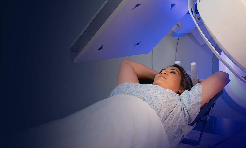
What is nuclear medicine?
Nuclear medicine uses very small amounts of radioactive materials, also called radiopharmaceuticals, to diagnose disease.
Radiopharmaceuticals are substances that are attracted to specific organs, bones or soft tissues. Where they gather, and how quickly they gather there, is the finding.
How the picture is made
The radiopharmaceuticals used in nuclear medicine emit gamma rays that can be detected externally by special types of cameras. These cameras work in conjunction with computers to form images that provide data about the area of the body being imaged.
How nuclear medicine differs from other scans
While X-rays image the body’s anatomy, nuclear medicine shows actual organ function and physiology.
It goes where it is needed
Radiopharmaceuticals are attracted to specific organs, bones or soft tissues. The tracer accumulates in the organ being examined.
It signals, the camera listens
The organ being examined emits radiation continuously during the exam. A special camera detects it, and a computer turns it into an image.
It answers what anatomy cannot
It allows clinicians to get medical information that would not be available otherwise, or that would require surgical intervention.
When is it used?
Nuclear medicine uses radioactive materials to help diagnose a variety of diseases and disorders, and to assess how different parts of your body are functioning.
-
01
Thyroid uptake and scans
Evaluating nodules, goiters, and hyperthyroidism.
-
02
Hepatobiliary (HIDA) scans
Evaluating gallbladder ejection fraction, biliary blockages, and liver function.
-
03
Whole-body bone scans
Detecting bone metastases, occult fractures, arthritis, and osteomyelitis.
-
04
Gastric emptying studies
Measuring stomach motility and digestive rate.
-
05
Myocardial perfusion imaging (MPI)
Assessing blood flow to heart muscle during rest and stress.
-
06
Pulmonary V/Q scans
Detecting pulmonary embolisms when CT contrast is contraindicated.
Inside the exam
The tracer goes in
You are given a small, safe amount of a radioactive tracer (a radiopharmaceutical), which may be injected intravenously, inhaled, or swallowed depending on the exam.
It finds its place
The tracer accumulates in the organ being examined. Depending on the exam, this is where the waiting comes in.
The camera listens
The organ being examined emits radiation continuously during the exam. That radiation is detected by a special camera and processed through a computer. You will be able to communicate with your technologist throughout.
A radiologist reads it
A contracted radiologist reviews your images and sends a report to your physician. Urgent findings are communicated right away.
How long the appointment takes
This is the one thing about nuclear medicine that surprises people, and it is worth knowing before you book. The scan is short. The tracer takes its own time.
Ask us how long your specific exam runs when you schedule, so you can plan the day around it rather than the other way around.
The tracer is given
Injected into a vein, inhaled, or swallowed, depending on the exam your physician ordered.
The tracer travels
The tracer accumulates in the organ being examined. Nothing is happening that you can feel, which is exactly the point.
You may be asked to come back
Depending on the exam, your technologist may ask you to return later, sometimes many hours later or the following day, to give the tracer time to distribute to the area of concern in your body.
The scan itself
You lie still while the camera collects the signal. The length of the exam varies, depending on type.
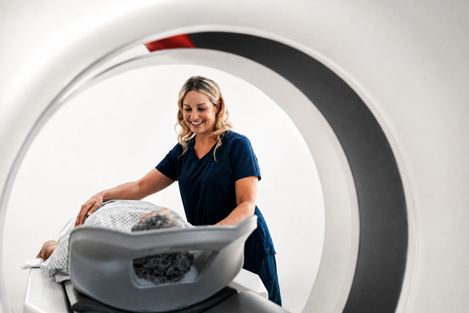
Is nuclear medicine safe?
Nuclear medicine is generally safe and well tolerated, using small amounts of radioactive material called radiotracers.
The radiation dose from a diagnostic nuclear medicine exam is typically low, though the amount varies depending on the type of exam. The lowest amount of radiation needed to obtain high-quality images is used.
Most patients experience few or no side effects from radiotracers. Allergic reactions are extremely rare and usually mild.
If you are pregnant, think you might be, or are breastfeeding, tell us when you schedule, as special precautions or instructions may be needed.
Exam preparation
We want your exam to be as comfortable and hassle-free as possible, so we provide all the information you will need to prepare before you arrive.
Your specific exam has its own instructions. You will find them on the Exam Preparation page.
All exam preparationWhat to bring
-
Prescription or referral from your physician.
-
A list of the medications you take, including non-prescription medications and supplements.
-
Insurance cards.
-
Any previous, relevant imaging exams and reports performed outside of our network, and your medical history.
Common questions
Nuclear medicine is generally safe and well tolerated, using small amounts of radioactive material called radiotracers. Most patients experience few or no side effects, and allergic reactions are extremely rare and usually mild.
While X-rays image the body’s anatomy, nuclear medicine shows actual organ function and physiology.
The radiation dose from a diagnostic nuclear medicine exam is typically low, though the amount varies depending on the type of exam. The lowest amount of radiation needed to obtain high-quality images is used.
You are given a small, safe amount of a radioactive tracer (a radiopharmaceutical), which may be injected intravenously, inhaled, or swallowed depending on the exam. The tracer accumulates in the organ being examined.
The length of the exam varies, depending on type. Depending on the exam, your technologist may ask you to return later, sometimes many hours later or the following day, to give the tracer time to distribute to the area of concern in your body.
Yes. You will be able to communicate with your technologist during the procedure.
Preparation depends on which nuclear medicine exam your physician ordered, and our staff will go through your instructions when you schedule. Two things apply to every exam: your dose is prepared specifically for your appointment time, so arriving on time is critical, and, unless your doctor told you to limit fluids, you should plan to drink extra fluids afterwards to help naturally flush the tracer from your system.

Find a nuclear medicine location near you
Nuclear medicine is not offered at every center. Confirm the exam you need when you book.