

+21.6%
Cancer detectionPowered by DeepHealth AI and Safeguard review.
2 million+
Mammograms annuallyAbout 5% of the United States total.


The power of AI behind your screening mammogram
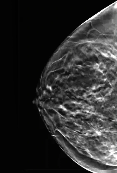
Without EBCD
The radiologist views the exam without assistance.
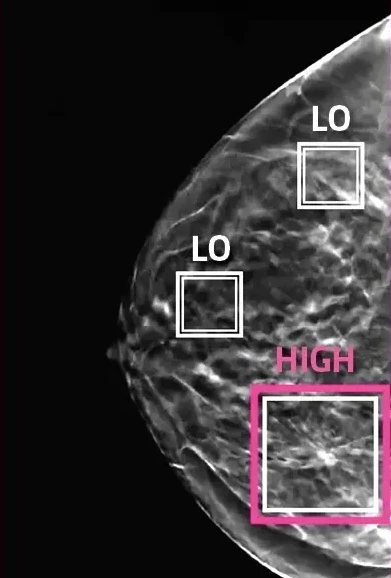
With EBCD
Suspicious areas are highlighted for the radiologist.
How Enhanced Breast Cancer Detection works
 01
After your exam, Artificial Intelligence (AI) technology is applied to your mammogram.
 02
On 3D mammograms, the AI marks areas of interest and how suspicious each looks, for the radiologist to review.
 03
After the radiologist reads your exam, the AI’s output is compared with their assessment.
 04
Certain exams with suspicious findings are reviewed by a second radiologist for determination of final results.
The ASSURE study
The benefits of RadNet’s EBCD (Enhanced Breast Cancer Detection) workflow, which leverages AI and rigorous clinical review to strengthen the effectiveness of mammography, are validated by the largest real-world analysis of AI-enabled breast cancer screening in the U.S. as of November 2025.
21.6%
Increase in cancer detection
The cancer detection rate rose from 4.6 to 5.6 per 1,000 screens after the AI workflow was added at 109 community imaging centers, compared with standard 3D mammography reading before it.
579K+
Mammograms evaluated
The ASSURE study evaluated more than 579,000 mammograms, making it the most comprehensive U.S. analysis of AI-enabled breast cancer screening as of November 2025.
22.7%
Higher detection in women with dense breasts
AI-enabled screening improved detection in dense-breast patients, addressing one of the most challenging limitations of mammography.
15%
Higher positive predictive value (PPV1)
Radiologists identified more true cancers per recall.
Equitable
Outcomes across patient populations
Study scale enabled statistical analyses by subpopulation, which revealed equitable performance across racial and ethnic groups, with no disparities widened.
Acceptable
No disproportionate increase in recalls
Recall rates rose modestly (10.6% to 11.1%) and stayed within ACR guidelines while detection and PPV1 improved.
Source: Louis LD, et al. Nature Health 1, 58–66 (2026). Increases are relative to standard 3D mammography reading before the AI workflow.
Through EBCD, RadNet has screened 1.5M+ women and helped detect 1,000+ additional cancers (as of November 2025).
Mammo Dx™ Artificial Intelligence, powered by DeepHealth. Mammo Dx is for 3D (tomosynthesis) screening mammograms only, not diagnostic mammograms. It is an adjunct tool, not intended to replace a radiologist’s own review of a mammogram — the final interpretation is made by a radiologist. Mammography does not detect all breast cancer lesions, and even with Mammo Dx some lesions may not be identified.
Comprehensive breast imaging
Breast imaging covers a range of advanced procedures radiologists use to detect and diagnose breast cancer. Mammography is where breast care starts — these are the exams we use alongside it, and who each one is for.
Mammography
An X-ray of the breast, and the exam most patients come to us for. It is used two ways — as a routine annual screen for women with no symptoms, and as a more detailed diagnostic study when there is something specific to look at.
- Screening mammography A routine X-ray of the breast used to detect breast cancer in women who have no signs or symptoms. It is a preventative measure, recommended annually from age 40 — or earlier for women considered high risk.
- Diagnostic mammography Performed when there are signs or symptoms of a potential breast issue — a lump, breast pain, nipple discharge, or a change found on a screening mammogram. It provides more detailed images to evaluate the area of concern closely and guide whether further testing is needed.
- 3D mammography (tomosynthesis) Images taken from several angles build a three-dimensional view of the breast, each scan taking about four seconds, so radiologists can examine the tissue layer by layer and pick up subtle abnormalities — particularly valuable for women with dense breast tissue. Software on our units removes the need for the extra images the exam once required, which keeps the dose low.
- What it feels like The breast is positioned and briefly compressed while each image is taken — usually two views of each breast. The compression is firm and lasts only a few seconds, and the whole exam runs 15–20 minutes.
Breast MRI
- What it is Powerful magnets and radio waves build very clear pictures of soft tissue — no radiation, and no compression of the breast. For the exams used to detect or evaluate cancer, contrast is given through an IV.
- Why it helps It finds small lesions that mammography sometimes misses.
- Who it is for Some women, because of family history, a genetic tendency or other risk factors, should be screened with MRI in addition to mammograms. Talk with your doctor about whether you should have additional tests.
Breast ultrasound
- What it is Sound waves instead of X-rays. A transducer is passed over the breast and the returning sound is translated into a picture on a monitor. It is generally painless, and with no radiation it carries very little risk.
- Why it helps It tells a fluid-filled cyst apart from a solid mass, and it reaches lesions close to the chest wall where mammography is less useful.
- Who it is for Women with dense breasts, and anyone whose mammogram or physical exam turned up something that needs a closer look.
Contrast-enhanced mammography (CEM)
- What it is A mammogram with contrast. You are given an IV, then a special mammogram captures two images within a second of each other — the ordinary picture, and one showing where the contrast has gone.
- Why it helps The contrast highlights areas of increased blood flow, which can make some cancers easier to see. It brings much of the sensitivity of MRI to a mammography platform.
- Who it is for Used as supplemental imaging for some women with dense breasts, and for those at intermediate risk.
Image-guided biopsies
When an abnormality requires tissue sampling, RadNet provides outpatient, minimally invasive image-guided biopsies. Procedures are performed under local anesthesia with no hospital stay.
| Biopsy type | Guidance | Primary indication | Technique |
|---|---|---|---|
| Stereotactic / 3D DBT biopsy | 3D digital mammography coordinates | Non-palpable microcalcifications or architectural distortions seen only on mammogram | Core needle samples target tissue through a tiny skin nick. |
| Ultrasound-guided biopsy | Real-time high-resolution ultrasound | Palpable lumps or solid masses visible on sonogram | Core needle retrieved under direct continuous visual tracking. |
| MRI-guided breast biopsy | High-field MRI guidance grid | Lesions visible strictly via breast MRI contrast enhancement | Core needle sampling performed inside a specialized grid localization. |
Care for patients at elevated risk
At select West Coast locations, when family history, a prior finding or a risk model points to elevated risk, RadNet coordinates what comes next — assessment, genetic testing and navigation — through our Breastlink breast care teams.
Integrated risk assessment
Screening patients are offered a no-cost high-risk questionnaire using the Tyrer-Cuzick model.
36-gene genetic testing panel
In partnership with Ambry Genetics and Cedars-Sinai Cancer, patients whose risk assessment meets NCCN criteria for genetic testing are offered comprehensive genetic screening for inherited cancer mutations.
Dedicated patient navigators
Individualized clinical guidance to coordinate diagnostic follow-up, surgical consultations and oncology referrals without delay.
Breastlink centers, where available
In California and Arizona, our Breastlink centers bring breast imaging, surgery and medical oncology together in a single practice. Visit Breastlink
Screening where you already are
In select markets, RadNet meets women where they live, work and shop to break down historical barriers to screening access.
Pure Mammography
Mammogram and ultrasound centers inside Roosevelt Field and Smith Haven malls on Long Island.
Available in New York.
Visit Pure MammographyMammogramNow at Super Walmart
Retail screening centers inside Super Walmart locations, reaching underserved populations and first-time screeners.
Available in California and Delaware.
Visit MammogramNowMobile mammography coaches
Mobile 3D mammography units servicing corporate campuses, rural communities and health fairs, with EBCD available at additional cost.
Available in select markets.
About mobile mammographyBreast imaging questions
The American College of Radiology recommends women aged 40 and older should have a screening mammogram every year and should continue to do so for as long as they are in good health.
No physician referral is required for screening mammography, but you must have an established healthcare provider who can receive and review your results.
EBCD pairs the radiologist with FDA-cleared AI and a built-in second review. After your exam, the AI marks areas of interest on your 3D mammogram and how suspicious each looks, then, after the radiologist reads the exam, the AI’s output is compared with their assessment — and where the radiologist reads the exam as normal but the AI flags high suspicion, a second breast specialist reviews it. Your mammogram experience remains the same: no additional radiation or time is required.
Yes — and it has been validated at scale. The ASSURE study, published in Nature Health, evaluated RadNet’s EBCD workflow across more than 579,000 mammograms, the largest real-world analysis of AI-enabled breast cancer screening in the U.S. as of November 2025. In that before-and-after comparison, the AI-augmented workflow had a 21.6% higher cancer detection rate (5.6 versus 4.6 per 1,000 screens) than standard 3D mammography reading.
Density is common, and it is not a diagnosis. In the ASSURE study, AI-enabled screening improved detection in dense-breast patients by 22.7% — addressing one of the most challenging limitations of mammography. Discuss what density means for your own screening with your physician.
Breast MRI is typically recommended for women with a lifetime risk of breast cancer of 20% or higher, including BRCA1/BRCA2 carriers and some women with a personal or strong family history, and for checking silicone breast implants.
Image-guided breast biopsies are outpatient, minimally invasive procedures performed under local anesthesia, with no hospital stay. The guidance method — 3D mammography, ultrasound or MRI — is matched to how the finding is best seen.
Yes. Please provide access to any mammograms performed at other facilities for vital year-over-year comparison. Without them your results may be delayed, or you may be asked to return for additional imaging.
Schedule your mammogram
No physician referral is required for screening mammography, but you must have an established healthcare provider who can receive and review your results.